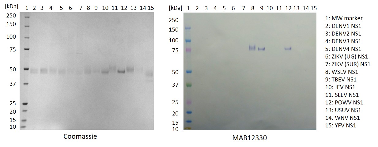
Western Blot: 100ng of each NS1 antigen was used for SDS-PAGE, in reduced form. Proteins were transferred using Transblot for 7 minutes at 25V. 5% dry milk in PBS-T was used as blocking buffer and dilution buffer for antibodies. Primary antibodies are given below, and goat anti-mouse-IgG-HRP secondary antibody (Biorad 103005) was used at 1:1000. All steps were carried out for 1h at room temperature with gentle rocking. KPL Membrane TMB was used for detection. Development time 30 seconds.
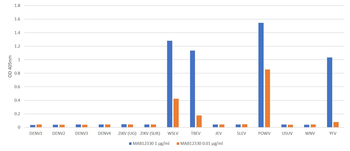
ELISA: All antigens coated at 0.5µg/ml in DPBS overnight at 2-8ᵒC. Plate washed 1 X 300µl/well TBS + 0.1% Tween20, blocked 300µl/well DPBS+1% BSA for an hour. Antibody diluted to 1.0µg/ml and 0.01µgml in DPBS + 1% BSA + 0.05% T20. Added at 100µl/well, incubated shaken 2h room temperature. Plate washed 3 X 300µl/well TBS-T wash buffer. Biorad goat anti-mouse IgG-HRP (103005) diluted 1 in 2500 in DPBS/1%BSA/0.05%T20, added at 100µl/well, incubated shaken 1h room temperature. Plate washed 6X 300µl/well TBS-T wash buffer. Europa TMB substrate added at 100µl/well and the plate developed for 2 min. static on the bench. Reaction stopped with 100µl/well 1M HCL and the plate was read within 4 min. at 405nm.
MOUSE ANTI-POWASSAN VIRUS NS1 ANTIBODY (M956)
Mouse monoclonal anti Powassan Virus NS1 antibody (clone M956) recognises NS1 protein from Powassan virus (POWV). The antibody is suitable for sandwich ELISA and suggested antibody pairs are; for capture, MAB12329, MAB12330 and MAB12331 paired with MAB12328, MAB12329, MAB12330 or MAB12331 for detection.
PRODUCT DETAILS – MOUSE ANTI-POWASSAN VIRUS NS1 ANTIBODY (M956)
- Mouse anti-Powassan virus NS1 (IgG1, clone M956). Recognises Powassan virus NS1 in ELISA.
- Antibody does not cross-react with NS1 from DENV, ZIKV, JEV, SLEV, USUV or WNV. It shows some cross-reactivity with WSLV, TBEV and YFV.
- Purified by Ion Exchange. >90% purity by SDS-PAGE.
- Presented in phosphate buffered saline, pH 7.2 with 0.05% sodium azide.
BACKGROUND
NS1 proteins are highly conserved structural proteins among the Flaviviridae family. Intracellular dimers of NS1 facilitate genome replication, whereas the secreted hexameric form plays a role in immunoevasion. Secreted NS1 has been identified as a potential diagnostic marker for early detection of infections. In addition to diagnostic potential, NS1 is being used in the development of therapeutics and NS1-based subunit vaccines (Rastogi, et al., 2016)
Powassan virus is a positive-sense, single-stranded RNA virus that belongs to the Flavivirus genus, of the Flaviviridae family. POWV is an arbovirus, transmitted by Dermacentor and Ixodes ticks depending on geography (Hicar et al., 2011). Infected ticks then transmit the virus to small-medium sized mammals, which act as reservoirs for POWV. The virus can also be transmitted to humans, which are an incidental, dead-end host (Hermance et al., 2017).
POWV is a member of the tick-borne encephalitis (TBEV) sero-complex of flaviviruses, and two genotypes, or lineages of POWV have been identified in Russia and the Western hemisphere. POWV is assigned to lineage I, and the Deer tick virus (DTV) to lineage II. Serologically, POWV and DTV are indistinguishable, and have both been linked to human disease, though show phylogenetic differences. Infection incidence by the POWV lineage have increased about 700% in the North American region over the last two decades (Fatmi et al., 2017). POWV is transmitted by the Ixodes scapularis tick, which is also the vector for Lyme disease and the expanding distribution of Ixodes and the high seroprevalence of DTV in small mammals in endemic regions suggests that POWV poses an increasing threat to public health.
First recognised as a human pathogen in 1958, POWV is the cause of a rare neuroinvasive disease. In humans, the incubation period varies between 1 – 5 weeks, and in many cases, individuals remain asymptomatic or may present with a febrile illness with mild fever, sore throat, headache, drowsiness and disorientation. Some of those infected can develop neurological complications, including encephalitis, aseptic meningitis and meningoencephalitis. Reports suggest that 10% of POWV cases are fatal, and that 50% of POWV survivors develop long-term neurological problems, including acute headaches, muscle wasting and memory problems (CDC).
REFERENCES
- Fatmi et al. (2017). Powassan Virus—A New Reemerging Tick-Borne Disease. Frontiers in Public Health. 5: 342.
- Hermance ME and Thangamani S. (2017). Powassan Virus: An Emerging Arbovirus of Public Health Concern in North America. Vector Borne Zoonotic Dis. 1; 17(7): 453–462.
- Hicar et al. (2011). Powassan Virus Infection Presenting As Acute Disseminated Encephalomyelitis In Tennessee. The Pediatric Infectious Disease Journal. 30(1):86-88
- Centers for Disease Prevention and Control (CDC): Powassan virus
- Rastogi et al. (2016). Flavivirus NS1: A Multifaceted Enigmatic Viral Protein. Virology Journal. 13:131.

")
 Powered by Bioz