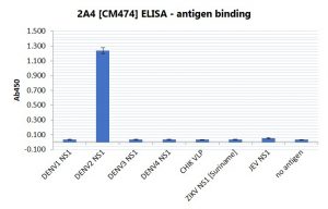
An ELISA plate was coated with 100ng of antigen per well, then blocked with 2% BSA. Primary antibody was used at a concentration of 1ug/ml, and the detection antibody used was Goat anti-mouse IgG:HRP (Bio-Rad, 1:2000). The substrate used was TMB (KPL).

Image by Virology Research Services Ltd
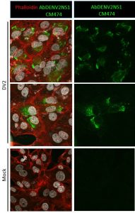
Immunofluorescence was carried out in Vero cells. Cells were seeded on coverslips and infected with DENV (serotypes 1, 2, 3 and 4) for 48h at MOI 1. Control coverslips consisted of uninfected Vero cells. After fixation with 4% PFA, samples were stained with Mouse anti-Dengue virus NS1 antibody (AbDENV2NS1-CM474). Antibody was diluted 1:500 and Triton X-100 was used as detergent. Imaging was performed using a Leica SP5 confocal microscope. Immunofluorescence was observed in DENV serotype 2 infected cells, stained with AbDENV2NS1-CM474, as shown above (Virology Research Services Ltd).
MOUSE ANTI-DENGUE VIRUS NS1 SEROTYPE 2 ANTIBODY (CM474)
Mouse anti Dengue virus NS1 antibody (serotype 2) is specific for the NS1 protein of Dengue virus serotype 2. It does not cross-react with Dengue NS1 from other serotypes, nor with NS1 from Zika virus or other flaviviruses. No cross-reactivity is seen with Chikungunya virus proteins (E1, E2 and C). This monoclonal mouse anti Dengue virus serotype 2 NS1 antibody is suitable for use in direct ELISA and in sandwich ELISA.
PRODUCT DETAILS – MOUSE ANTI-DENGUE VIRUS NS1 SEROTYPE 2 ANTIBODY (CM474)
-
- Mouse anti-Dengue virus seroytpe 2 NS1 monoclonal antibody (clone CM474).
- Greater than 95% purity by SDS-PAGE and buffered in PBS, pH7.4.
BACKGROUND
Dengue is caused by one of four different serotypes of small RNA viruses in the family Flaviviridae, DENV1–4. NS1 is a major non-structural protein expressed by the Dengue virus. The NS1 monomer is a glycosylated protein of approximately 45kD, which associates with lipids and forms a homodimer inside infected cells. It is necessary for viral replication, and is also secreted into the extracellular space as a hexameric lipoprotein particle, which is involved in immune evasion and pathogenesis by interacting with components from both the innate and adaptive immune systems, as well as other host factors. NS1 is one of the major antigenic markers for viral infection with Dengue. It is secreted from infected cells and is found in serum at detectable levels that overlap with peak viremia and RNA detection. This also coincides with the start of detectable IgM in acute primary cases and IgG in acute nonprimary cases. Elevated levels of serum NS1 directly indicates increased viral burden and indicates a positive correlation between viremia and NS1 profiles (Ambrose et al., 2017).
Dengue fever is ranked by the World Health Organization (WHO) as the most critical mosquito-borne viral disease, globally. More than 40 percent of the world’s population, in more than 100 countries are at risk of dengue infection. The most significant dengue epidemics in recent years have occurred in Southeast Asia, the Americas and the Western Pacific. Each year, an estimated 390 million dengue infections occur around the world. Of these, 500,000 cases develop into dengue haemorrhagic fever – a more severe form of the disease, which results in up to 25,000 deaths annually.
With more than one-third of the world’s population living in areas at risk of transmission, dengue infection is a leading cause of illness and death in the tropics and subtropics (WHO for Guidelines for Diagnosis, Treatment, Prevention & Control, 2009) As many as 100 million people are infected yearly. Dengue is caused by any one of four related viruses transmitted by mosquitoes. There are no vaccines available to prevent infection with dengue virus.


