
 Powered by Bioz
DIPHTHERIA TOXIN
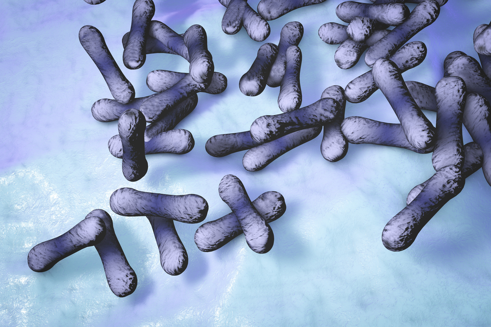
Diphtheria toxin manufactured by The Native Antigen Company is a native protein produced by culture of Corynebacterium diphtheriae strain NCTC 10648 in optimal growth medium, followed by harvest, concentration, and purification through several rounds of chromatography. This results in a toxin > 98% pure by SDS-PAGE and of high quality and minimal lot-to-lot variation, suitable for IVD assays, cellular research and in vivo work. It is offered in 1mg vial size and the toxin is lyophilised for ease of transportation and storage. Each batch is tested for activity using an in-vitro CHO cytotoxicity assay, demonstrating 100% cell death at a concentration of 10ngml.
PRODUCT DETAILS – DIPHTHERIA TOXIN
- 58kDa native toxin from Corynebacterium diphtheriae strain NCTC 10648.
- Greater than 98% purity by SDS-PAGE.
- Each batch is activity tested for performance against a reference lot (MKCK6150).
- Lyophilised in 0.01 M Tris and 0.001 M Na2EDTA, 0.05% D-lactose, pH 7.5.
BACKGROUND
Diphtheria is a disease caused by Diphtheria toxin (DT), a potent exotoxin that is a member of the family of ADP-ribosylating bacterial toxins. DT is secreted by strains of the gram-positive bacterium Corynebacterium diphtheria carrying a lysogenic bacteriophage, which contains the toxin gene (Freeman, 1952). The toxin is secreted as a single polypeptide chain of 535 amino acids (Mwt 58.342) containing two major functional domains, known as domains A and B. The amino terminal A domain (1-193 amino acids) carries the catalytic domain for ADP-ribosylation of elongation factor 2 (EF2). The carboxyl terminal B domain (194-535 amino acids) is further divided into receptor (R) binding and transmembrane (T) domains (Choe, 1992). The B domain promotes binding of DT to cells, the internalization of DT by receptor-mediated endocytosis and the entry of the A polypeptide chain into the cytosolic compartment (Greenfield, 1983).
In humans, DT binds to epidermal growth factor receptor (HB-EGFR) on the surface of cells, and the toxin–receptor complex undergoes receptor-mediated endocytosis. The arginine-rich segment connecting the A and B domains is then cleaved to yield active DT-A and DT-B fragments. The A fragment is translocated across the endocytic membrane into the cytoplasm, a process that is facilitated by DT-B. In the cytoplasm, the catalytic domain A inactivates elongation factor-2, which is an essential component of the protein translation machinery, halting protein synthesis and causing cell death (Collier, 1975).
Since its discovery in 1888, Diphtheria toxin has become one of the most extensively studied toxin molecules. In humans, DT is a potent toxin and the mechanism of DT toxicity has been well documented.However, rodent cells do not have the correct form of EGFR to bind DT and are resistant to the toxic effects of extracellular DT. This has enabled scientists to generate transgenic mouse models that specifically express DT receptor (DTR) in restricted cell populations, making them sensitive to extracellular DT and subsequent cell death.
Similar transgenic mouse models have been generated to analyse the role of FoxP3 regulatory T-cells in vivo. Regulatory T-cells (T-regs) maintain self-tolerance and express the forkhead box transcription factor Foxp3, which is essential for their development and function. Selective depletion of FoxP3 T-reg cells provides insight into the role of T-regs in the immunopathology of autoimmune disease, transplantation, cancer and infectious disease. To deplete FoxP3 T-reg cells, investigators generated bacterial artificial chromosome (BAC) transgenic mice, named ‘depletion of regulatory T-cell’ (DEREG) mice, which expressed a DTR enhanced green fluorescent protein (eGFP) under the control of the Foxp3 locus. Foxp3 T-reg cells in newborn mice are then be ablated by injection of DT. The DEREG mouse model has enabled scientists to clearly identify and study the effects of FoxP3 T-reg cells in vivo, which has been difficult using existing antibody based methods (Lahl, 2007).
REFERENCES
- Freeman VJ, Morse IU. (1952). Further observations on the change to virulence of bacteriophage-infected a virulent strains of Corynebacterium diphtheria. J Bacteriol. Mar;63(3):407-14.
- Choe S, Bennett MJ, Fujii G, Curmi PM, Kantardjieff KA, Collier RJ, Eisenberg D. (1992). The crystal structure of diphtheria toxin. Nature. May 21;357(6375):216-22.
- Greenfield L, Bjorn MJ, Horn G, Fong D, Buck GA, Collier RJ, Kaplan DA. (1983). Nucleotide sequence of the structural gene for diphtheria toxin carried by corynebacteriophage beta. Proc Natl Acad Sci U S A. Nov;80(22):6853-7.
- Collier RJ. (1975). Diphtheria toxin: mode of action and structure. Bacteriol Rev. Mar;39(1):54-85.
- Lahl K, Loddenkemper C, Drouin C, Freyer J, Arnason J, Eberl G, Hamann A, Wagner H, Huehn J, Sparwasser T. (2007). Selective depletion of Foxp3 regulatory T cells induces a scurfy-like disease. J Exp Med. Jan 22;204(1):57-63.