
Canine Parvovirus
Canine parvovirus or ‘parvo’ is a highly contagious disease of dogs and causes acute gastrointestinal illness, particularly in puppies and adolescent dogs. It is caused by canine parvovirus (CPV-2) which first emerged among dogs in Europe in 1976 and by 1978 had spread worldwide causing an epidemic of myocarditis and inflammation in the intestines (gastroenteritis) of dogs. CPV is closely related to feline panleukopenia virus (FPV) and probably arose as the result of a small number of genetic mutations in FPV that allowed it to expand its host range to infect dogs. The original CPV-2 has since evolved into two antigenic variants, CPV-2a and CPV-2b, which have progressively replaced it; more recently a new antigenic variant, CPV-2c, has also appeared. There are vaccines available against CPV, but due to its maintenance in nature in wild canids, its ease of transmission and its ability to mutate rapidly there is a need for ongoing epidemiological research and surveillance.
The Native Antigen Company offers recombinant Canine Parvovirus virus-like particles and antibodies, in support of research into CPV biology and infection.
Canine Parvovirus Background
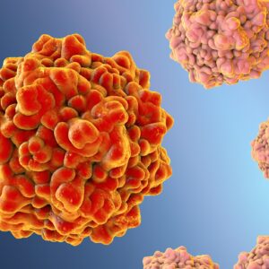
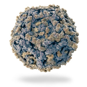
Canine Parvovirus (CPV-2) is a non-enveloped, single-stranded DNA virus, in the Parvoviridae family with icosahedral symmetry (20-26 nm in diameter). CPV is a relatively new disease that was first recognized in 1978 and then rapidly spread worldwide, becoming the most important enteric virus infecting canids (Carmichael, 2005).
There are two types of canine parvovirus, canine minute virus (CPV-1) and CPV-2. CPV-2 causes the most serious disease and affects domesticated dogs and wild canids, including foxes, wolves and skunks (Holmes et al., 2013). There are variants of CPV-2 called CPV-2a and CPV-2b, identified in 1979 and 1984 respectively (Shackelton et al., 2005) and more recently CPV-2c, a Glu-426 mutant, discovered in Italy, Vietnam, and Spain (Decaro et al., 2006). Canine Parvovirus (CPV-2) has 98% identity at the amino-acid level to feline panleukopenia (also a parvovirus), differing only in two amino acids in the viral capsid protein VP2 (Carter & Wise, 2004). Phylogenetic analysis showed that all CPVs derived from a single common ancestor, and that the strains were mostly similar to viruses from different wildlife animals. CPV-2 likely arose when it acquired mutations that allowed binding to the canine transferrin receptor (TfR) type-1. TfR plays a key role in the susceptibility of cells to infection by parvoviruses (reviewed in Miranda & Thompson, 2016). The original CPV-2 rapidly evolved into two antigenic variants, CPV-2a and CPV-2b, which progressively replaced the original CPV-2 (reviewed in Nandi & Kumar, 2010). More recently a new antigenic variant, CPV2c, has appeared (reviewed in Nandi & Kumar, 2010). The prevalence of CPV-2b (VP2 426Asp) has been reported in different countries within Europe, Asia, Africa, the Americas and Oceania and was found to be the predominant antigenic variant in Ireland, the UK, the USA, African and Asian countries. Both CPV-2a and -2b were distributed in equal proportion in Belgium, Switzerland and Austria and a sequence with VP2426Asp was also isolated from a dog in Russia (reviewed in Miranda & Thompson, 2016).
Canine Parvovirus (CPV-2) is highly contagious and is transmitted by the faecal-oral route through contact with faeces from infected dogs or contaminated surfaces. Dogs that develop the disease show signs of the illness within 3 to 7 days, including lethargy, vomiting, fever, and diarrhoea (usually bloody). Following ingestion, the virus replicates in the lymphoid tissue in the throat, and then spreads to the bloodstream. From there, the virus infects cells in the lymph nodes, intestinal crypts, and the bone marrow. There is depletion of lymphocytes in lymph nodes and necrosis and destruction of the intestinal crypts. Anaerobic bacteria that normally reside in the intestines can then cross into the bloodstream, a process known as translocation, with bacteremia leading to sepsis. The most common bacteria involved in severe cases are Clostridium, Campylobacter and Salmonella species. This can lead to a syndrome known as systemic inflammatory response syndrome (SIRS) and a range of complications such as hypercoagulability of the blood, endotoxaemia and acute respiratory distress syndrome (ARDS). Bacterial myocarditis has also been reported secondarily to sepsis. Three to four days following infection, the virus is shed in the faeces for up to three weeks, and the dog may remain an asymptomatic carrier and shed the virus periodically (Ettinger et al., 1995). In the more common, less severe form, mortality is ~10 percent; but without treatment, dogs with severe disease can die within 48-72 hours and mortality can reach 91%.
The main method for controlling the disease in domestic animals is by vaccination. However, given the ability of Canine Parvovirus (CPV-2) for rapid evolution, the continued need for epidemiological surveillance and improvement of vaccines is likely to be a necessity (reviewed in Miranda & Thompson, 2016).
References
- Carmichael L (2005). An annotated historical account of canine parvovirus. J. Vet. Med. B Infect. Dis. Vet. Public Health. 52 (7–8): 303–11.
- Carter, G.R. & Wise, D.J. (2004). Parvoviridae. A Concise Review of Veterinary Virology. IVIS.
- Decaro et al. (2006). First detection of canine parvovirus type 2c in pups with haemorrhagic enteritis in Spain. J. Vet. Med. B Infect. Dis. Vet. Public Health. 53 (10): 468–72.
- Ettinger, Stephen J.; Feldman, Edward C. (1995). Textbook of Veterinary Internal Medicine (4th ed.). W.B. Saunders Company.
- Holmes et al. (2013). Frequent Cross-Species Transmission of Parvoviruses among Diverse Carnivore Hosts. Journal of Virology. 87 (4): 2342–2347.
- Miranda & Thompson (2016). Canine parvovirus: the worldwide occurrence of antigenic variants. J Gen Virol. 97(9):2043-2057.
- Nandi & Kumar (2010). Canine Parvovirus: Current Perspective. Indian J Virol. 21(1): 31–44.
- Shackelton et al. (2005). High rate of viral evolution associated with the emergence of carnivore parvovirus. Proc. Natl. Acad. Sci. USA. 102 (2): 379–384.
Canine Parvovirus Antigens
CPV-2 VP2 is produced in the form of virus-like particles (VLP) in insect cell lines using state-of-the-art expression techniques which yield VLPs of exceptional quality and purity.
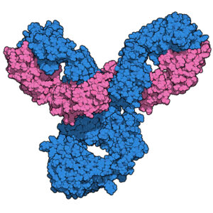
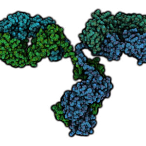
Canine Parvovirus Antibodies
The Native Antigen Company is pleased to offer a range of five antibodies against our CPV-2 VP2 antigen. These antibodies have been validated for use in sandwich ELISA and other applications, allowing you to develop diagnostic kits for epidemiological studies and vaccine research.
Questions?
Check out our FAQ section for answers to the most frequently asked questions about our website and company.