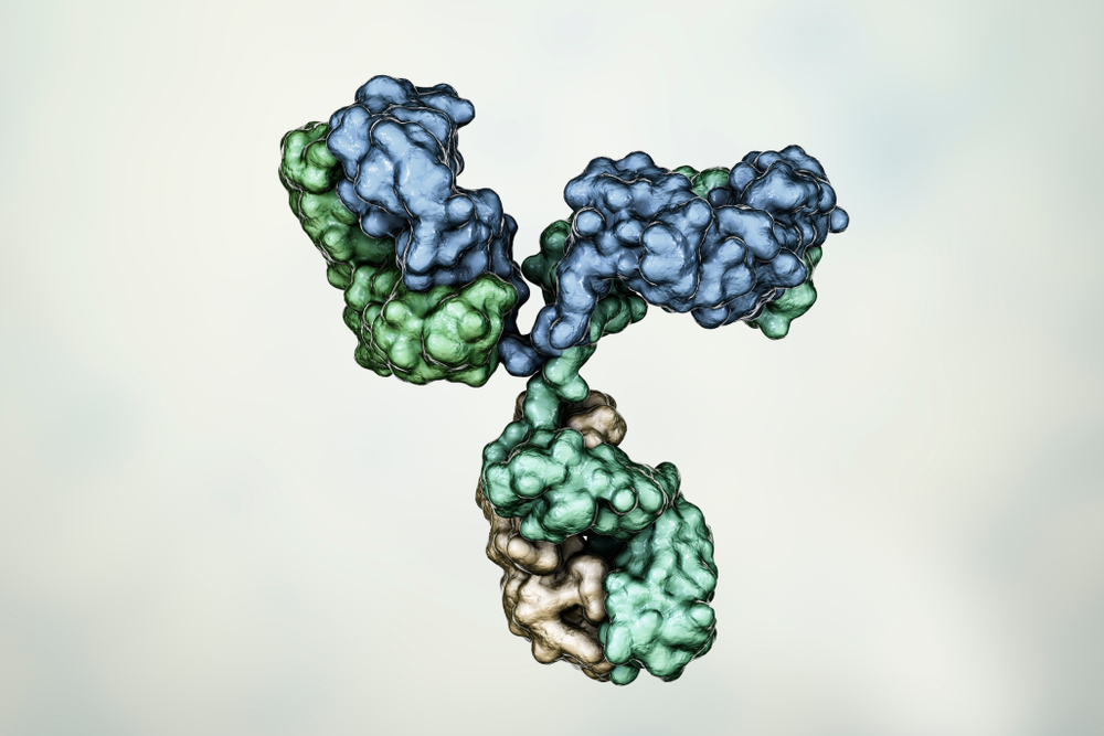
MOUSE ANTI-FELINE LEUKEMIA VIRUS P27 ANTIBODY (7226)
Mouse anti Feline leukemia virus p27 antibody (7226) is specific for the p27 of FeLV. The antibody is suitable for enzyme immunoassay (EIA) applications (ELISA and Lateral Flow).
PRODUCT DETAILS – MOUSE ANTI-FELINE LEUKEMIA VIRUS P27 ANTIBODY (7226)
- Mouse anti Feline leukemia virus p27 monoclonal antibody (7226).
- Specific for FeLV p27.
- Suitable for use in IFA, ELISA and Lateral Flow applications.
- Purified by Protein G Sepharose chromatography.
- Presented in phosphate buffered saline, pH 7.2, 0.1% sodium azide.
BACKGROUND
Feline leukemia virus (FeLV) is a widespread pathogen of the domestic cat and (Cornell University, 2016) and was first described in 1964. It is an RNA virus in the subfamily Oncovirinae belonging to the Retroviridae family. The virus comprises 5′ and 3′ LTRs and three genes: Gag (structural), Pol (enzymes) and Env (envelope and transmembrane); the total genome is ~9.6 kbp. Following synthesis of a DNA copy of the viral RNA genome, it integrates into the genome of the target cell as a provirus. This provirus remains in the genome of the cell for the life span of the cell and, upon cellular division, the viruses bud from the membrane of the infected cell, undergoing the final phase of maturation with cleavage of the Pr65 group antigen (Gag) precursor into the mature structural proteins p15 matrix (MA), p27 capsid (CA) and p10 nucleocapsid (NC).
FeLV assembles at the plasma membrane. The precursor of the viral structural proteins Pr65Gag is thought to attach to the underside of the membrane. Once bound, the Pr65Gag proteins multimerise, triggering the membrane to bend around the forming core. Env proteins associate with the nascent particle through their co-localisation on the membrane until an immature particle is formed. A scission event then takes place, releasing the immature virion, at which time the viral protease cleaves Pr65Gag into distinct matrix (MA), capsid (CA) and nucleocapsid (NC) proteins. As they bud from the infected cell, nascent virions acquire the envelope glycoprotein Env, comprising the surface (SU) glycoprotein gp70 and transmembrane (TM) protein p15E. Gp70 is the target for neutralising antibodies in recovered cats and is an essential component of FeLV vaccines (Willett and Hosie, 2013).
The prevalence of FeLV in cats has decreased significantly in the past 25 years since the development of an effective vaccine and more accurate testing procedures (Cornell University, 2016). The majority of in-practice tests for FeLV detect viral antigen (capsid protein p27) in blood, plasma or serum. P27 is the most abundant viral protein in the plasma of viraemic cats. Its utility as a diagnostic marker for FeLV viraemia is only possible because cats do not appear to respond serologically to the protein, an observation that has led to speculation that cats may be largely immunologically tolerant to p27 through exposure to endogenously expressed FeLV Gag (Willett and Hosie, 2013).
REFERENCES
- Feline leukemia virus factsheet. Cornell University College of Veterinary Medicine, May 2016.
- Feline leukaemia virus factsheet. Small Animal Veterinary Surveillance Network (SAVSNET), University of Liverpool, September 2019.
- Willett and Hosie (2013). Feline leukaemia virus: half a century since its discovery. Vet J. 195(1):16-23.

