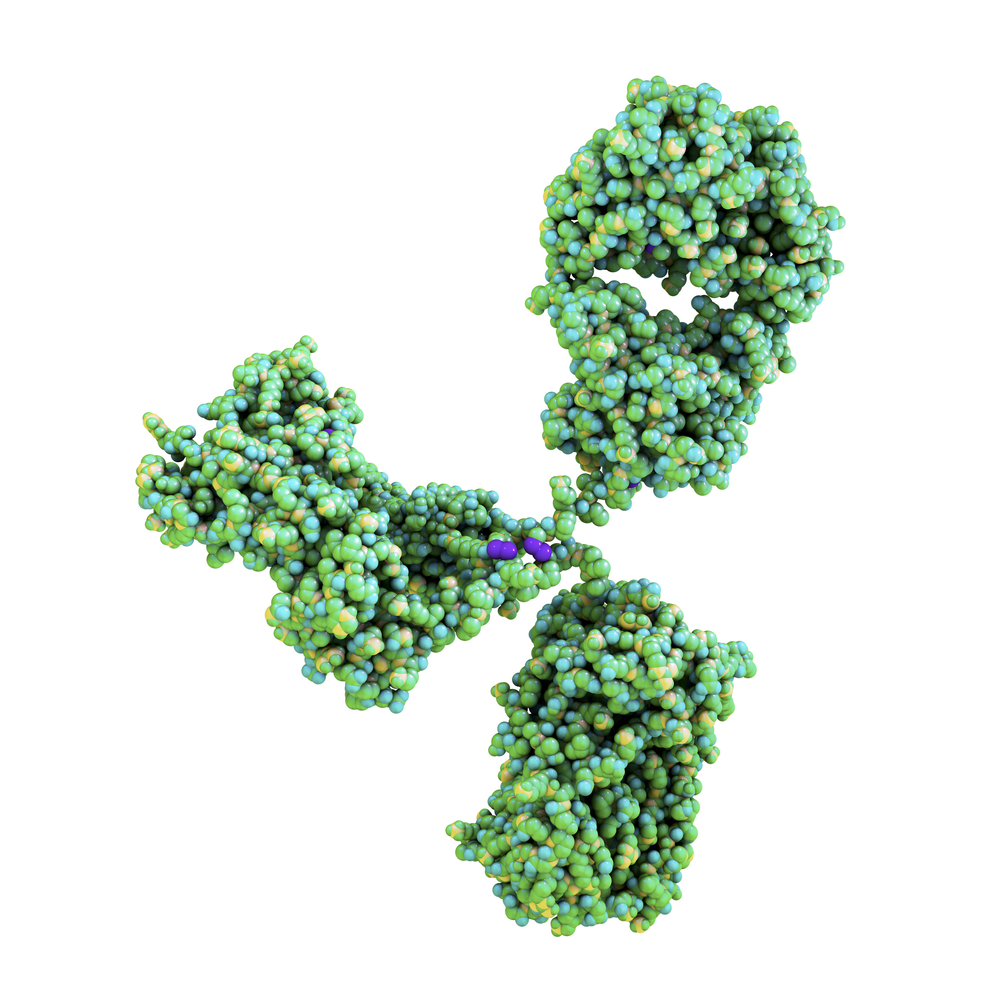
MOUSE ANTI-DIPHTHERIA TOXIN ANTIBODY (7801)
Mouse anti Diphtheria Toxin antibody is specific for intact diphtheria toxin and toxoid. The Diphtheria Toxin antibody does not react with free A or B subunits. Diphtheria Toxin antibody is suitable for use in ELISA assays with mouse anti diphtheria toxin antibody (MAB12178).
PRODUCT DETAILS – MOUSE ANTI-DIPHTHERIA TOXIN ANTIBODY (7801)
- Mouse anti-Diptheria toxin monoclonal IgG1 antibody (clone 7801).
- Greater than 90% purity by SDS-PAGE and buffered in PBS, pH7.2.
BACKGROUND
Diphtheria toxin (DT) is a potent exotoxin that is a member of the family of ADP-ribosylating bacterial toxins. It was first identified in 1888 as the causative agent of an infectious disease in humans known as diphtheria. DT is secreted by strains of the gram-positive bacterium Corynebacterium diphtheria carrying a lysogenic bacteriophage, which contains the toxin gene (Freeman, VJ).
The toxin is secreted as a precursor molecule and cleaved to produce a single polypeptide chain of 535 amino acids (Mwt 58.342) containing two major functional domains, known as domains A and B. The amino terminal A domain (1-193 amino acids) carries the catalytic domain for ADP-ribosylation of elongation factor 2 (EF2). The carboxyl terminal B domain (194-535 amino acids) is further divided into receptor (R) binding and transmembrane (T) domains (Choe, S). The B domain promotes binding of DT to cells, the internalization of DT by receptor-mediated endocytosis and the entry of the A polypeptide chain into the cytosolic compartment (Greenfield, L).
In humans, DT binds to heparin-binding epidermal growth factor receptor (HB-EGFR) on the surface of cells, and the toxin–receptor complex undergoes receptor-mediated endocytosis. The arginine-rich segment connecting the A and B domains is then cleaved to yield active DT-A and DT-B fragments. The A fragment is translocated across the endocytic membrane into the cytoplasm, a process that is facilitated by DT-B. In the cytoplasm, the catalytic domain A inactivates elongation factor-2, which is an essential component of the protein translation machinery, halting protein synthesis and causing cell death (Collier, RJ).
REFERENCES
- Freeman, V.J. et al (1952). Further observations on the change to virulence of bacteriophage-infected a virulent strains of Corynebacterium diphtheria. J Bacteriol. Mar;63(3):407-14.
- Choe, S. et al. (1992). The crystal structure of diphtheria toxin. Nature. May 21;357(6375):216-22.
- Greenfield, L. et al. (1983). Nucleotide sequence of the structural gene for diphtheria toxin carried by corynebacteriophage beta. Proc Natl Acad Sci U S A. Nov;80(22):6853-7.
- Collier, R.J. (1975). Diphtheria toxin: mode of action and structure. Bacteriol Rev. Mar;39(1):54-85.

