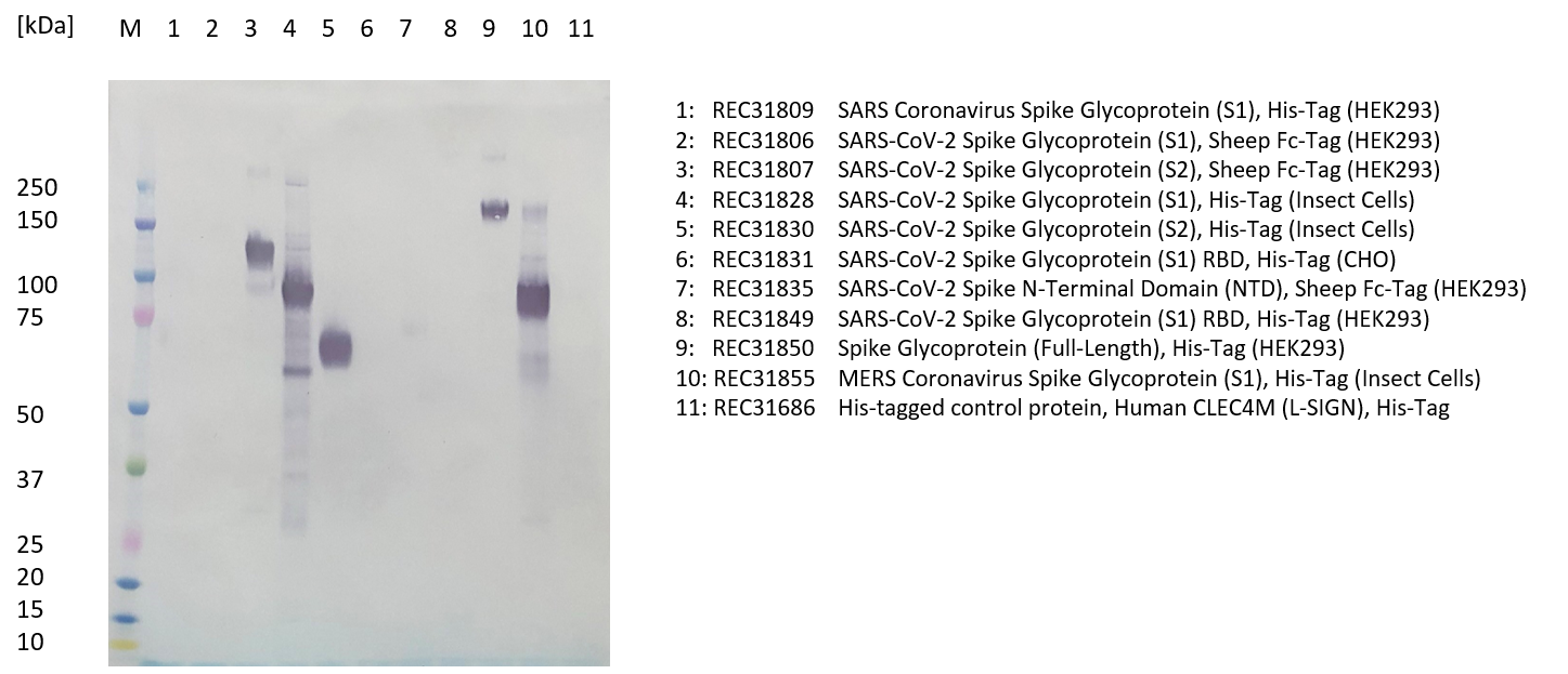
Western blot: Recombinant protein (100ng) was loaded into each lane and separated by SDS-PAGE in a Novex 4-12% Bis-Tris gel at 200V. Western blot was carried out using rabbit anti SARS-CoV-2 Spike (S2) polyclonal antibody (1mg/ml, 1:1000) and STAR124P secondary antibody (1mg/ml, 1:1000).
Antibody detected SARS-CoV-2 spike S2 subunit (lanes 3 and 5), and full-length spike protein (lane 9). It did not detect SARS-CoV-2 spike S1 subunit (lane 2 and 4), receptor binding domain (RBD) (lanes 6 and 8), spike N-terminal domain (NTD) (lane 7), SARS-CoV spike S1 subunit (lane 1) or control protein (lane 11). A band was visible for insect-cell expressed spike subunit 1 and insect-cell expressed MERS-CoV S1, likely due to the presence of host-cell proteins (antigen was purified from baculovirus-infected insect cells).

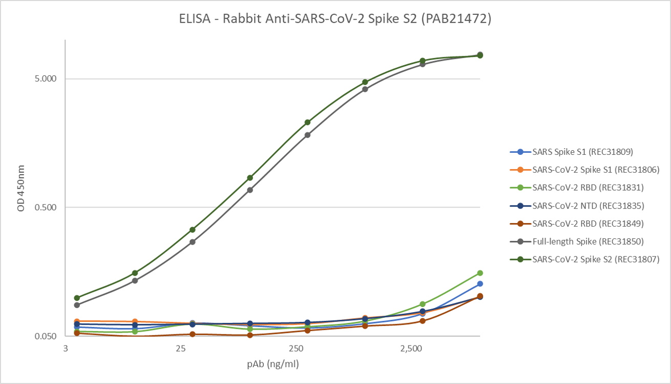
ELISA: Plates coated overnight at 2-8ºC in DPBS, antigen (1.0µg/ml) in 100µl/well. Blocked 2 x 300µl/well DPBS + 1% BSA, soaked 1 hour. Washed 1 X 300µl/well PBS + 0.1% Tween 20. Dilutions of antibody (100µl/well) applied in DPBS/1% BSA/0.1% Tween 20. Incubated shaken 3 hours 23ºC. Washed 3 X 300µl/well PBS + 0.1% Tween 20. Anti-rabbit Ig-HRP (Sigma A0545, 1:10,000) in DPBS/1% BSA/0.1% Tween 20, 100µl/well. Incubated shaken 3 hours, 23ºC. Washed 6 X 300µl/well PBS + 0.1% Tween 20. TMB (Europa MO701A) added (100µl/well), 15 min static incubation. Stopped (200µl 1M HCl) and read at 450nm. ODs out of range estimated from OD 405nm reading.
Antibody recognises SARS-CoV-2 Spike S2 subunit and full-length spike. It does not cross-react with SARS-CoV-2 Spike S1 subunit, receptor binding domain (RBD), or N-terminal domain (NTD).
RABBIT IgG ANTI-SARS-COV-2 SPIKE (S2) POLYCLONAL ANTIBODY
SARS-CoV-2 Spike (S2) polyclonal antibody is an antibody that recognizes the SARS-CoV-2 Spike glycoprotein, subunit 2 (S2) and is suitable for use in ELISA and WB. Antibody does not cross-react in ELISA with HCoV-229E full-length spike protein (REC31880).
PRODUCT DETAILS – RABBIT IgG ANTI-SARS-COV-2 SPIKE (S2) POLYCLONAL ANTIBODY
- SARS-CoV-2 Spike (S2) polyclonal antibody binds the S2 domain of the SARS-CoV-2 Spike protein.
- This antibody was made by immunising rabbits with a recombinant SARS-CoV-2 Spike subunit 2 antigen (REC31830).
- Protein A affinity purified from culture supernatant.
- Suitable for use in ELISA and WB.
BACKGROUND
SARS-CoV-2 is part of the Coronaviridae family, subfamily Orthocoronavirinaes and includes the genera Alphacoronvirus, Betacoronavirus, Gammacoronavirus, and Deltacoronavirus. Alphacoronaviruses and betacoronaviruses typically infect mammals, whereas gammacoronoviruses and deltacoronaviruses generally infect avian species (and sometimes mammals). Two types of alphacoronaviruses (229E and NL63) and two types of betacoronaviruses (OC43 and HKU1) are pathogens of humans and cause the common cold. Whereas, SARS-CoV-1, the Middle Eastern respiratory syndrome coronavirus (MERS-CoV), and SARS-CoV-2 (all betacoronaviruses) cause more severe disease in humans including, respiratory, gastrointestinal, hepatic, and neurologic diseases (Amanat & Krammer, 2020).
SARS-CoV-2 has a large single-stranded positive-sense RNA genome encoding for several open reading frames. One frame encodes the spike protein (S protein), a class I fusion protein that forms homotrimers protruding from the viral surface and mediates coronaviruses attachment and adhesion to human target cells via a host receptor. Spike has two different functional regions, S1 and S2. The S1 subunit consists of four core domains, S1A to S1D. The distal S1 domain mediates receptor association and stabilization, whereas the S2 domain promotes structural rearrangements and membrane fusion. Coronaviruses use different regions of S1 domain to interact with specific binding receptors. For SARS-CoV-2, the receptor binding domain (RBD) interacts with the peptidase domain of the angiotensin-converting enzyme 2 (ACE2) receptor from bats, civet cats, swine, cats, ferrets, non-human primates (NHPs), and humans (Amanat & Krammer, 2020). ACE2 is a membrane-anchored carboxypeptidase highly expressed by airway epithelial and type I and II alveolar epithelial cells (Perrotta et al., 2020).
REFERENCES
- Amanat F, Krammer F. SARS-CoV-2 Vaccines: Status Report. Immunity. 2020;52(4):583-589.
- Perrotta F, Matera MG, Cazzola M, Bianco A. Severe respiratory SARS-CoV2 infection: Does ACE2 receptor matter?. Respir Med. 2020;168:105996.

 Powered by Bioz