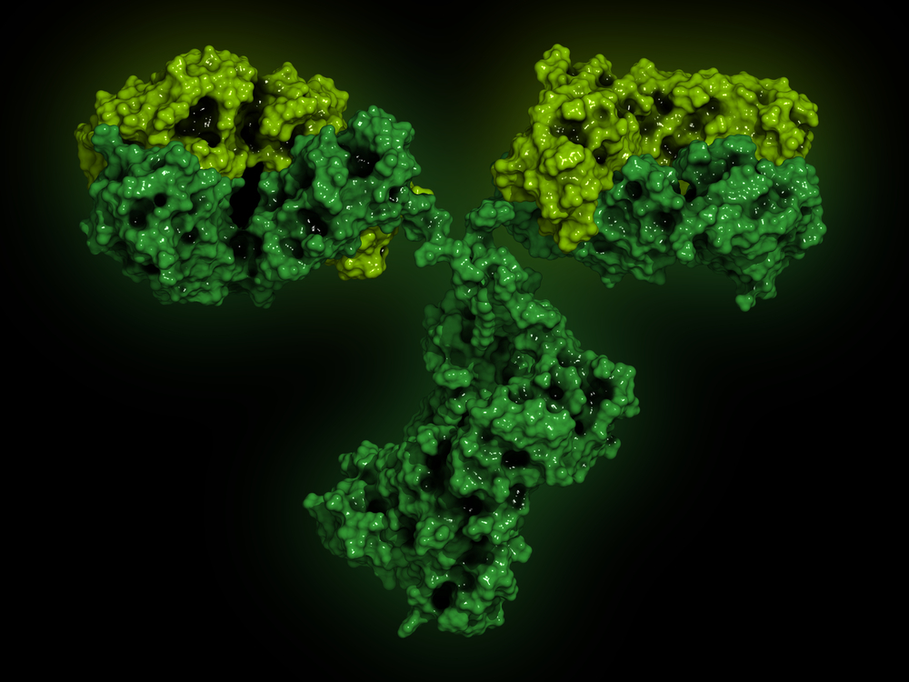
Mouse monoclonal antibody specific for Chikungunya virus VLP (clone 16A12)
Mouse monoclonal antibody specific for Chikungunya virus VLP (clone 16A12) recognizes the E2 protein when presented within the VLP or live virus. It demonstrates negligible cross-reactivity with other members of the alphavirus family, including Western Eastern and Venezuelan Encephalitis viruses. There is no cross-reactivity to Zika, Dengue or other flavivrius antigens.
This antibody is suitable for use in ELISA, Western Blot (non-denatured samples) and in immunofluorescence. The antibody has also been reported to neutralise virus in PRNT assays.
PRODUCT DETAILS – Mouse monoclonal antibody specific for Chikungunya virus VLP (clone 16A12)
- Monoclonal IgG1 antibody (clone 16A12).
- Greater than 95% purity by SDS-PAGE and buffered in PBS, pH7.4.
- Suitable for the development of highly specific immunoassays.
BACKGROUND
Chikungunya virus is the aetiological agent of chikungunya fever. CHIKV belongs to the Alphavirus genus, and is an enveloped, single-stranded positive-sense RNA virus (Strauss & Strauss, 1994). The alphavirus genome encodes four non-structural proteins (nsP1 to nsP4) and five structural proteins (capsid, E3, E2, 6K and E1).
CHIKV is transmitted to humans by Aedes mosquitoes, and disease is characterized by a rapid onset of fever, myalgia and often a rash (usually maculopapular), with chronic disease characterized by episodic, and often debilitating, polyarthralgia/polyarthritis. (Suhrbier et al., 2012). The largest epidemic of CHIKV disease ever reported began in 2004 and has since been responsible for up to 6.5 million human cases, primarily in Africa and Asia, with imported cases reported in over 40 countries. CHIKV infection is symptomatically similar to infection with Zika virus and Dengue virus, and differential diagnosis using immunoassay based testing is important in patient management.
E2, the receptor binding protein, contains residues critical for immunogenicity, host range, and tissue/cell tropism (Voss et al, 2010). The E2 protein consists of three domains (A, B, and C), of which A and B have been found to contain the majority of residues that affect cell attachment and or tissue/cell tropism.

