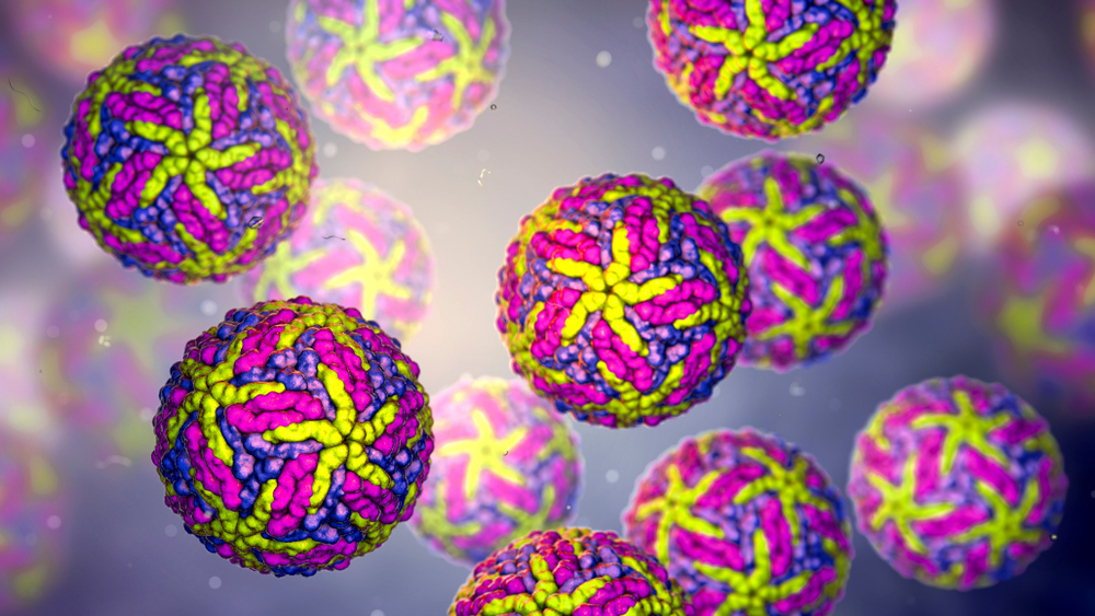
RHINOVIRUS TYPE 1A LYSATE
Rhinovirus type 1A lysate produced from MRC-5 or HeLa cells. Purified using sucrose density gradient ultracentrifugation, heat-inactivated and tested for absence of viral growth in validate tissue culture infectivity assays.
PRODUCT DETAILS – RHINOVIRUS TYPE 1A VIRUS LYSATE
- Purified rhinovirus type 1A lysate.
- Purified by sucrose density gradient ultracentrifugation.
- Heat-inactivated and verified using validated tissue culture infectivity assays.
- Appropriate for the development of immunoassays, Western blotting, dot blotting and other protein-based assays.
BACKGROUND
Human rhinoviruses (HRVs) were first discovered in the 1950s in an effort to identify the causative agent of the common cold (Jacobs et al., 2013). HRVs are positive-sense, single-stranded RNA viruses of the family Picornaviridae, genus Enteroviridae. HRVs were once classified into only two species, HRV-A and -B, according to phylogenetic sequence criteria. However, testing of clinical specimens in the 1960/70s yielded around 100 different strains, which were subsequently serotyped (Palmenberg et al., 2010). This led to the division of the original 99 strains into two the species HRV-A (containing 74 serotypes) and HRV-B (containing 25 serotypes). The HRV genome is translated into a single polyprotein product that is cleaved by virally-encoded proteases to produce 11 proteins: capsid proteins VP4, VP2, VP3, VP1 and non-structural proteins 2A, 2B, 2C, 3A, 3B, 3C and 3D. The VP1-3 proteins are responsible for the antigenic diversity of HRVs, while VP4 acts to anchor viral RNA to the capsid. There are 60 copies of the 4 structural proteins in each virion which produce an icosahedral structure (Jacobs et al., 2013). While previously thought to cause only relatively benign infections of the upper respiratory tract, HRVs have now been found to be associated with chronic pulmonary disease, asthma and severe bronchiolitis (Gutman et al., 2007).
REFERENCES
- Gutman, JA. et al. (2007). Rhinovirus as a Cause of Fatal Lower Respiratory Tract Infection in Adult Stem Cell Transplantation Patients: A Report of Two Cases. Bone Marrow Transplantation. 40(8):809.
- Jacobs, SE. et al. (2013). Human Rhinoviruses. Clinical Microbiology Reviews. 26(1): 135-162.
- Palmenberg, AC. et al. (2010). Analysis of the Complete Genome Sequences of Human Rhinovirus. Journal of Allergy and Clinical Immunology. 125(6):1190-1199.

