 Powered by Bioz
RABBIT ANTI-BORRELIA BURGDORFERI SENSU STRICTO (B31) ERPN/OSPE ANTIBODY
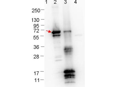
Rabbit anti-Borrelia burgdorferi ErpN/OspE polyclonal antibody, prepared against ErpN (OspE/F-Related Protein D), also known as Arp37, from the spirochete B. burgdorferi, for use in ELISA and western blotting applications.
PRODUCT DETAILS – RABBIT ANTI-BORRELIA BURGDORFERI SENSU STRICTO (B31) ERPN/OSPE ANTIBODY
- Rabbit anti-B. burgdorferi sensu stricto ErpN/OspE polyclonal IgG antibody (strain B31).
- Greater than 95% purity by SDS-PAGE and buffered in 0.02 M Potassium Phosphate, 0.15 M Sodium Chloride, pH 7.2.
BACKGROUND
Strain B31 is the type strain (ATCC 35210) for this organism and was derived by limited dilutional cloning from the original Lyme-disease tick isolate obtained by A. Barbour (Johnson, et al., 1984). Several studies have demonstrated that infected humans and animals produce antibodies directed against Erp proteins within the first 2-4 weeks of infection, suggesting Erp synthesis occurs during the initial stages of vertebrate infection. Surface-exposed Erp proteins may then facilitate interactions with host tissues during the establishment of vertebrate infection (Fraser, et al., 1997).
Erp proteins from B. burgdorferi are believed to be lipoproteins, based on their predicted amino acid sequences. The spirochete migrates from the tick midgut during feeding to its salivary glands and are thus transmitted to the mammal host. This transition may be facilitated by changes in expression of some B. burgdorferi genes. It is believed that expression of the various proteins associated with the spirochete may be regulated by the changes in tick life cycle, changes in conditions during tick feeding (such as temperature, pH, and nutrients) and/or in coordination with the course of infection of the mammal host (Stevenson, et al., 1996).
REFERENCES
- Fraser, C. M. et al., 1997. Genomic sequence of a Lyme disease spirochaete, Borrelia burgdorferi. Nature, Volume 390, pp. 580-586.
- Johnson, R.C., et al. 1984. Borrelia burgdorferi sp. nov.: etiologic agent of Lyme disease. Int J Syst Bacteriol, 34, pp. 496–497.
- Stevenson, B., Tilly, K. & Rosa, P. A., 1996. A Family of Genes Located on Four Separate 32-Kilobase Circular Plasmids in Borrelia burgdorferi B31. J Bacteriol, 178(12), pp. 3508-3516.