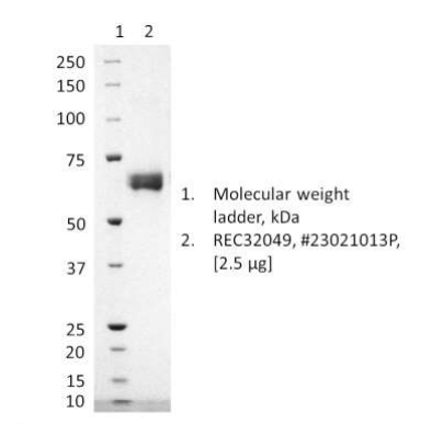
SDS-PAGE: Coomassie-stained reducing SDS-page showing purified : Human parainfluenzavirus 3 haemagglutinin-neuraminidase protein (REC32049), batch 23021013P.
Human parainfluenzavirus 3 haemagglutinin-neuraminidase (HN) protein, C-terminal His-tag
Price range: $915.56 through $3,890.19 excl. VAT
Recombinant human Parainfluenza Virus Type 3 haemagglutinin-neuraminidase (HN) protein, manufactured in HEK293 cells.
Human parainfluenza virus 3 haemagglutinin-neuraminidase (HN) protein, C-terminal His-tag
Recombinant human Parainfluenza Virus Type 3 haemagglutinin-neuraminidase (HN) protein, manufactured in HEK293 cells.
PRODUCT DETAILS – HUMAN PARAINFLUENZA VIRUS TYPE 3 HN PROTEIN (HEK293), C-TERMINAL HIS-TAG
- Purified Human Parainfluenza Virus Type 3 haemagglutinin-neuraminidase (HN) protein, strain Wash/47885/57, NCBI Accession number AAA46846.1
- Manufactured in mammalian HEK293 cells.
- Purified by immonbilised metal affinity chromatography, followed by ion exchange chromatography.
- Presented in 20mM sodium phosphate pH7.0, 210mM NaCl at >90% purity.
BACKGROUND
Human parainfluenza viruses (HPIV) belong to a diverse group of enveloped single-stranded RNA viruses within the Paramyxoviridae family, a large and expanding group of viruses that includes metapneumovirus and the mumps, measles and respiratory syncytial viruses. Paramyxoviruses are enveloped animal viruses with non-segmented negative-strand RNA genomes (complementary to mRNA), which are identified almost exclusively in nucleocapsid structures. The HPIV genome comprises approximately 15,000 nucleotides and encodes six common structural proteins: ‘large’ (L) nucleocapsid protein, P and N, which are closely associated with viral RNA, haemagglutinin-neuraminidase (HN), and fusion (F) and membrane (M) proteins (reviewed in Pawełczyk & Kowalski, 2017). HPIVs bind and replicate in the ciliated epithelial cells of the upper and lower respiratory tract and the extent of the infection correlates with the location involved (reviewed in Branche & Falsey, 2016). Human parainfluenza virus type 3 enters cells by directly fusing with the cell membrane. During entry, the viral surface glycoproteins HN, a receptor-binding protein, and F cooperate to mediate fusion upon receptor binding (Xu et al., 2013). The HN and fusion glycoproteins are the major targets for neutralizing antibodies (reviewed in Branche & Falsey, 2016). F is a class I viral fusion protein and has at least 3 conformational states: pre-fusion native state, pre-hairpin intermediate state, and post-fusion hairpin state. During viral and plasma cell membrane fusion, the heptad repeat (HR) regions assume a trimer-of-hairpins structure, positioning the fusion peptide in close proximity to the C-terminal region of the ectodomain. The formation of this structure appears to drive fusion of viral and plasma cell membranes and directs fusion of viral and cellular membranes leading to delivery of the nucleocapsid into the cytoplasm.
Human rhinovirus (HRV), respiratory syncytial virus (RSV), influenza virus and coronaviruses are considered the most important viral respiratory pathogens. However, seasonal HPIV epidemics result in a significant burden of disease in children and account for 40% of pediatric hospitalizations for lower respiratory tract illnesses (LRTIs) and 75% of croup cases. A wide spectrum of illness including colds, croup, bronchiolitis, and pneumonia are attributed to these ubiquitous pathogens. The most severe disease is found among immunocompromised patients and treatment at present remains largely supportive. Viral coinfections may also be a cause of pneumonia, including coinfection with both HPIV and human coronaviruses such as HCoV-229E (Gonzales Zamora, 2018). Several promising antiviral drugs are in development and are in early-stage clinical trials. Continued research for new vaccines and therapeutics is therefore needed (reviewed in Branche & Falsey, 2016).
REFERENCES
- Branche AR, Falsey AR. Parainfluenza Virus Infection. Semin Respir Crit Care Med. 2016;37(4):538-554.
- Gonzales Zamora, J. A. (2018). Pneumonia Caused by Coronavirus 229E and Parainfluenza 3 Coinfection in a Lung Transplant Recipient. Infectious Diseases in Clinical Practice, 26(1), e3–e4.
- Pawełczyk M, Kowalski ML. The Role of Human Parainfluenza Virus Infections in the Immunopathology of the Respiratory Tract. Curr Allergy Asthma Rep. 2017;17(3):16.
- Xu R, Palmer SG, Porotto M, et al. Interaction between the hemagglutinin-neuraminidase and fusion glycoproteins of human parainfluenza virus type III regulates viral growth in vivo. mBio. 2013;4(5):e00803-e813.


