
 Powered by Bioz
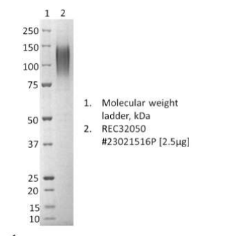
SDS-PAGE: Coomassie-stained reducing SDS-page showing purified Marburg GP1 (REC32050), batch : 23021516P

Marburg virus Glycoprotein GP1, C-terminal His-tag (HEK293)
Price range: £502.68 through £2,509.80 excl. VAT
Recombinant Marburg GP1 (Musoke Kenya, 1980) produced in HEK293 cells. Marburg virus Glycoprotein 1, C-terminal His-tag has been developed for use in immunoassay and vaccine development.
Marburg virus Glycoprotein GP1, C-terminal His-tag (HEK293)
Marburg GP1 (Musoke Kenya, 1980) produced in HEK293 cells. Marburg virus GP1, C-terminal His-tag has been developed for use in immunoassay and vaccine development.
PRODUCT DETAILS – MARBURG VIRUS GLYCOPROTEIN (GP1) (MUSOKE KENYA, 1980)
- Recombinant Marburg virus GP1 was expressed in HEK293 cells, and contains a C-terminal His-tag.
- Sequence strain is Marburg virus/Musoke Kenya, 1980 (MARV-Mus), NCBI Accession Number: AAR85456.1
- Protein was purified from culture supernatant by a series of immobilized metal affinity chromatography, followed by ion exchange chromatography and dialysis
- Presented as Liquid, in DPBS
BACKGROUND
Marburg virus (MARV) is a hemorrhagic fever virus of the Filoviridae family and a member of the species Marburg marburgvirus, genus Marburgvirus. MARV causes a form of viral hemorrhagic fever in humans and nonhuman primates and is considered to be extremely dangerous. The virus can be transmitted by exposure to Old World fruit bats or it can be transmitted between people via body fluids.
The GP gene is the fourth gene along the genome of every filovirus (Sanchez et al., 1993) and encodes a trans-membrane protein GP localized at the virus surface. Marburg virus contains only one transmembrane protein (GP) which is responsible for receptor recognition on target cells, and triggers viral attachment and entry. GP, a type I membrane protein of approximately 220 kDa, is acylated and highly glycosylated carrying N- and O-linked sugar side chains. GP is transported through the exocytotic pathway toward the plasma membrane where budding of virions takes place. In the trans-Golgi network, GP is proteolytically activated by the prohormone convertase furin into two subunits GP(1) and GP(2) (Sänger et al., 2002). The GP gene also codes for additional proteins, whose functions are not yet fully understood (Martin et al., 2016).
Viral cell entry is a critical point for infection, and as such, this step is targeted for the design of antiviral molecules. Antibodies have demonstrated that inhibitors targeting filovirus entry may provide a potent antiviral strategy (Martin et al., 2016).
REFERENCES
- Martin B, Hoenen T, Canard B, Decroly E. Filovirus proteins for antiviral drug discovery: A structure/function analysis of surface glycoproteins and virus entry. Antiviral Res. 2016 Nov;135:1-14.
- Sanchez A, Kiley MP, Holloway BP, Auperin DD. Sequence analysis of the Ebola virus genome: organization, genetic elements, and comparison with the genome of Marburg virus. Virus Res. 1993 Sep;29(3):215-40.
- Sänger C, Mühlberger E, Lötfering B, Klenk HD, Becker S. The Marburg virus surface protein GP is phosphorylated at its ectodomain. Virology. 2002 Mar;295(1):20-29.