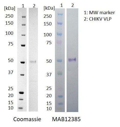
Western Blot: 100ng of antigen was used for SDS-PAGE, in reduced form. Proteins were transferred using Transblot for 7 min. at 25V. 5% dry milk in PBS-T was used as blocking buffer and dilution buffer for antibodies. Primary antibodies and goat anti-mouse-IgG-HRP secondary antibody (Biorad 103005) were used at 1:1000. All steps were carried out for 1h at room temperature with gentle rocking. KPL Membrane TMB was used for detection. Development time 30 sec.
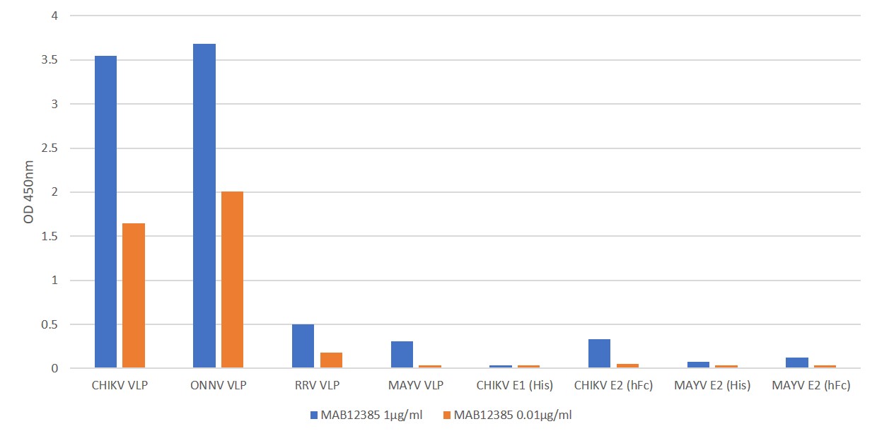
ELISA: Coat plate with 0.5µg/ml of antigens in 1X DPBS overnight at 2-8 degrees. Block for 1.5 h using 1%BSA/DPBS.300µl/well. Wash plate 3X using Tris wash buffer. Add 100µl of prepared antibody dilutions. Incubate for 2 h at RT at 800rpm. Wash plate 3X using Tris wash buffer. Add 100µl of Goat anti mouse IgG HRP antibody (1:5000 dilution) to all the wells and incubate for 1 h at RT,800rpm. Wash plate 4X using Tris wash buffer. Add 100µl HKTMB and incubate for ~2 min. at RT. Add 100µl 1M HCl to stop the reaction and read the plate at 450nm.
MOUSE ANTI-CHIKUNGUNYA VIRUS VLP ANTIBODY (D7)
This mouse anti Chikungunya virus VLP antibody (D7) recognises Chikungunya virus-like particles (VLP) containing E1, E2 and Capsid proteins.
PRODUCT DETAILS – MOUSE ANTI-CHIKUNGUNYA VIRUS VLP ANTIBODY (D7)
- Mouse anti Chikungunya virus VLP antibody recognises the Chikungunya virus VLP, containing E1, E2 and capsid proteins.
- Antibody also cross-reacts with O’nyong’nyong virus VLPs in ELISA.
- Antibody does not cross-react in ELISA with CHIKV envelope proteins (E1 and E2), Mayaro virus (MAYV) VLPs, MAYV E2 or Ross River virus (RRV) VLPs in ELISA.
- Presented in PBS with 0.02% Proclin 300.
BACKGROUND
Chikungunya virus is a member of the genus Alphavirus in the family Togaviridae. Chikungunya fever is a mosquito-borne disease first identified in Tanzania in 1953. Since 2004 there have been extensive outbreaks in Africa and Asia, and in 2013 the first cases were identified in the Caribbean, and by September 2014 more than 650,000 cases had been reported in the Americas. There is a risk that the virus will be imported to new areas by infected travellers.
Chikungunya virus is spread to people by the bite of an infected mosquito. Chikungunya fever usually starts 2–4 days after chikungunya virus infection and the most common symptoms of infection are fever and joint pain. Other symptoms may include headache, muscle pain, joint swelling, or rash.
Laboratory diagnosis is generally accomplished by testing serum or plasma to detect virus, viral nucleic acid, or virus-specific immunoglobulin (Ig) M and neutralizing antibodies. Viral culture may detect virus in the first 3 days of illness; however, chikungunya virus should be handled under biosafety level (BSL) 3 conditions. During the first 8 days of illness, chikungunya viral RNA can often be identified in serum. Chikungunya virus antibodies normally develop toward the end of the first week of illness. Therefore, to definitively rule out the diagnosis, convalescent-phase samples should be obtained from patients whose acute-phase samples test negative (CDC, 2019).
Diagnosis of CHIKV may be hampered by the fact that clinical symptoms of CHIKV are similar to those seen in cases of Dengue virus and Zika virus infection. Therefore, differential diagnosis is an important consideration in areas where flaviviruses such as Dengue and Zika co-circulate. Currently, there is no specific treatment available for the treatment of Chikungunya fever or licensed vaccine for the prevention of CHIKV infection.
Virus-Like Particles (VLPs) are an emerging vaccine technology and consist of protein shells comprising outer proteins specific to the virus in question. The Native Antigen Company’s Chikungunya virus VLP is composed of E1, E2 and capsid proteins, from Chikungunya virus sequences. Concentration and purification are then performed by a series of ultracentrifugation and chromatographical methods which result in VLPs of exceptional quality and purity. These antigens are then used to produce antibodies with higher affinity and greater sensitivity, which can used for multiple applications.
REFERENCES


 Powered by Bioz