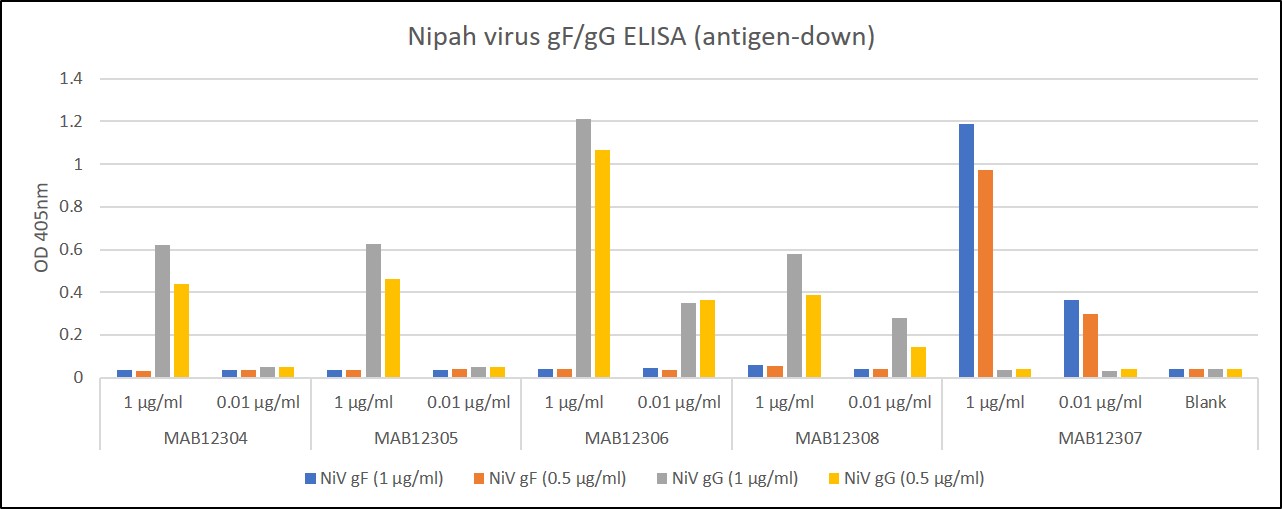
ELISA: Nipah virus Glycoprotein F (gF) and Glycoprotein G (gG) antigens were coated at 0.5µg/ml and 1µg/ml at RT in DPBS for an hour. Plate washed 1 X 300µl/well TBS + 0.1% Tween20, blocked 300µl/well DPBS+1% BSA 1h at room temperature. Detection antibodies diluted to 1.0µg/ml and 0.01µgml in DPBS + 1% BSA + 0.05% T20. Added at 100ul/well, incubated shaken 2h room temperature. Plate washed 3 X 300µl/well TBS-T wash buffer. Biorad goat anti-mouse IgG-HRP secondary antibody (103005) diluted 1 in 2500 in DPBS/1%BSA/0.05%T20, added at 100µl/well, incubated shaken 1h room temperature. Plate washed 6X 300µl/well TBS-T wash buffer. Detection with KPL/Seracare SureBlue TMB substrate added at 100µl/well and the plate developed for 3 min. static on the bench. Reaction stopped with 100µl/well 1M HCL and the plate was read within 5 min. at 405nm. MAB12304, MAB12305, MAB12306 and MAB12308 are specific to NiV gG. MAB12307 is specific to NiV gF.
MOUSE ANTI-NIPAH VIRUS GLYCOPROTEIN F ANTIBODY (CG11)
Mouse anti Nipah virus (NiV) glycoprotein G monoclonal antibody is specific for the gF protein of NiV.
PRODUCT DETAILS – MOUSE ANTI-NIPAH VIRUS GLYCOPROTEIN F ANTIBODY (CG11)
- Mouse IgG monoclonal antibody (clone CG11-B1-D6-G3).
- Specific for NiV gF protein. It shows no cross reactivity with gG protein in antigen down ELISA.
- Immunized using pool of recombinant NiV glycoproteins G and F.
- Purified from hybridoma cell culture supernatant by affinity chromatography on Protein G. Final buffer is PBS, pH7.4.
BACKGROUND
Nipah virus (NiV) is an enveloped single stranded negative sense RNA virus that belongs to the Henipavirus genus, which is a new member of the Paramyxoviridae family. Nipah infection was first recognised in Malaysia 1998/1999, where a major NiV outbreak occurred in pigs and humans. A subsequent outbreak of NiV in Singapore also pointed to pigs as an intermediate host. However, outbreaks in India and Bangladesh did not. The natural host for NiV has now been identified as the fruit bat, of the Pteropus genus, with swine acting as intermediate host in some cases. Reports suggest that transmission of Nipah virus to humans can occur through contact with NiV infected bats, food contaminated by bat’s excrement, infected pigs and other NiV infected humans.
Nipah, the disease caused by NiV infection is now endemic in South Asia and several outbreaks of NiV infection have been reported in India and Bangladesh. The symptoms of Nipah virus infection in humans can include rapidly developing fever, abdominal pain, nausea, vomiting, acute respiratory syndrome and severe encephalitis, which is fatal in a high percentage of cases (WHO). In 2015, the World Health Organization highlighted NiV infection as an emerging disease requiring accelerated R&D to advance in vitro diagnostic development, vaccine design and therapeutics (WHO, 2015).
REFERENCES

 Powered by Bioz