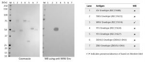
Western Blot: WB using anti-West Nile virus (WNV) Envelope protein antibody (MAB12297): 100ng of antigen in reducing sample buffer was loaded onto each lane (see table above). SDS-PAGE and Western blot (WB) was conducted. Membranes were incubated for 1h in Blocking Buffer (5% Dry Milk powder in PBS-T). Membranes were then incubated with PAB21464 antibody at 1µg/ml in Blocking Buffer for 2h at room temperature. Membranes were washed 3x with PBS-T for 5 minutes, then incubated for 1h at room temperature in Blocking Buffer containing a 1:2,500 dilution of STAR124P (Bio-Rad). Membranes were then washed 3x for 5 minutes with PBS-T, then developed using TMB Membrane substrate (KPL).
Rabbit Anti-West Nile Virus Envelope Protein Antibody (9E2)
Price range: $1,360.03 through $2,017.96 excl. VAT
This rabbit monoclonal antibody (9E2) binds to protein E, between amino acids 321 and 466, of a range of WNV strains, including Hp-94, Vlg99-27889, A-72, Vlg00-27924, Tur-2914, Eg101 and A-1640. This antibody has been tested in western blot and ELISA on JEV, TBEV, YFV, DENV and Zika envelope proteins, and shown to have no cross-reactivity. Antibody has a neutralizing effect in an in vitro model of WNV.
RABBIT ANTI-WEST NILE VIRUS ENVELOPE PROTEIN ANTIBODY (9E2)
Rabbit anti-West Nile Virus Envelope protein antibody (9E2) binds to protein E, between amino acids 321 and 466, of a range of WNV strains, and has been tested in Western blot and ELISA on JEV, TBEV, YFV, DENV and Zika Envelope proteins, and shown to have no cross-reactivity. Antibody has a neutralizing effect in an in vitro model of WNV.
PRODUCT DETAILS – RABBIT ANTI-WEST NILE VIRUS ENVELOPE PROTEIN ANTIBODY (9E2)
- Rabbit anti-West Nile virus Envelope protein antibody (9E2).
- WNV virions (Vlg99-27889) used as immunogen and antibody purified by Protein A affinity chromatography.
- Antibody binds to protein E, between amino acids 321 and 466, of a range of WNV strains, including Hp-94, Vlg99-27889, A-72, Vlg00-27924, Tur-2914, Eg101 and A-1640.
- Antibody has a neutralizing effect in an in vitro model of WNV.
- Presented in PBS with 0.02% Proclin 300.
BACKGROUND
West Nile Virus (WNV) is a member of the flavivirus genus and belongs to the Japanese encephalitis antigenic complex of the family Flaviviridae. It is commonly found in Africa, Europe, the Middle East, North America and West Asia. It is the leading cause of mosquito-borne disease in the continental United States and is most commonly spread to people by the bite of an infected mosquito (birds are its natural host). Approximately 20% of people who are infected will show symptoms of West Nile fever, including fever, headache, tiredness, and body aches, nausea, vomiting, occasionally with a skin rash (on the trunk of the body) and swollen lymph glands. Severe West Nile disease (neuroinvasive disease) can be fatal, with headache, high fever, neck stiffness, stupor, disorientation, coma, tremors, convulsions, muscle weakness, and paralysis. It is estimated that approximately 1 in 150 persons infected with the West Nile virus will develop a more severe form of disease.
West Nile virus can be diagnosed by IgG and IgM ELISA, RT-PCR and virus isolation by cell culture. Although a vaccine is available for horses, there is no vaccine for humans.


