LISTERIA MONOCYTOGENES CELLS, HEAT-INACTIVATED
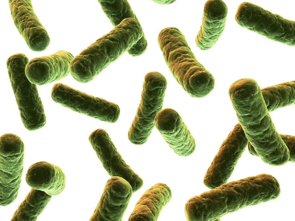
Heat-killed Listeria monocytogenes cells in dextran solution. Antigen is intended for use as a positive control in immunoassay development for Listeria detection.
PRODUCT DETAILS – LISTERIA MONOCYTOGENES CELLS, HEAT-INACTIVATED
- Heat-killed Listeria monocytogenes cells, genus specific in dextran solution.
- Part of the BacTrace® range of antigens and antibodies.
- This product is ideally suited for use as a positive control in immunoassays designed for the detection of Listeria. It provides verification of the functionality of the assay system.
- Product is considered non-hazardous as defined by The Hazard Communication Standard (29 CFR 1910.1200).
BACKGROUND
Listeria is a food-borne pathogen responsible for a disease called listeriosis, which is potentially lethal in immunocompromised individuals. It is a genus of bacteria that acts as an intracellular parasite in mammals and as of 2020 was known to contain 21 species. Listeria species are Gram-positive, rod-shaped, and facultatively anaerobic, and do not produce endospores. The major human pathogen in the genus Listeria is L. monocytogenes. It is usually the causative agent of the relatively rare bacterial disease listeriosis, an infection caused by eating food contaminated with the bacteria. Listeriosis can cause serious illness in pregnant women, newborns, adults with weakened immune systems and the elderly, and may cause gastroenteritis in others who have been severely infected (Radoshevich & Cossart, 2018).
Listeriosis is a serious disease for humans; the overt form of the disease has a case-fatality rate of around 20%. The two main clinical manifestations are sepsis and meningitis. Meningitis is often complicated by encephalitis, when it is known as meningoencephalitis, a pathology that is unusual for bacterial infections.
L. ivanovii is a pathogen of mammals, specifically ruminants, and has rarely caused listeriosis in humans.
REFERENCES
- Radoshevich L, Cossart P. Listeria monocytogenes: towards a complete picture of its physiology and pathogenesis. Nat Rev Microbiol. 2018 Jan;16(1):32-46.


