
 Powered by Bioz
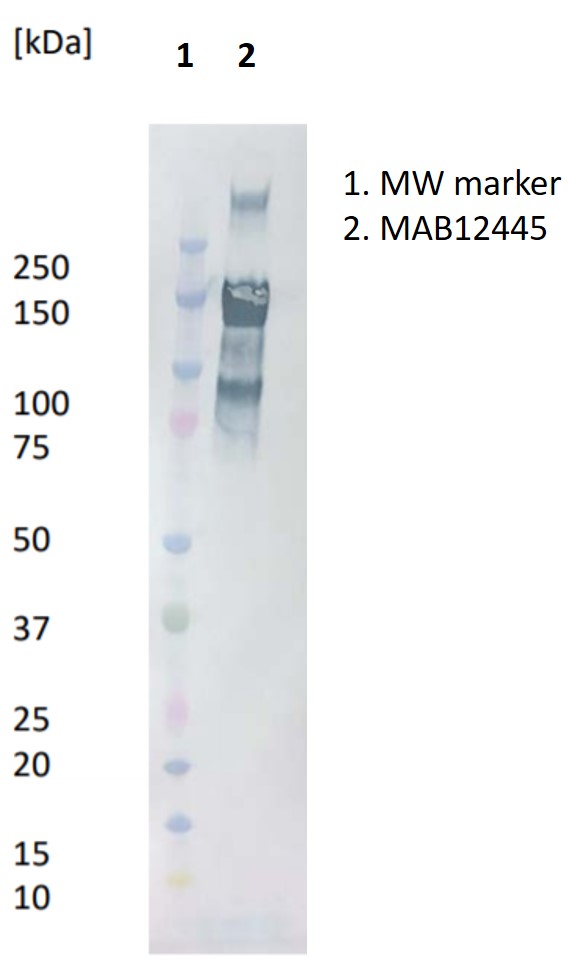
Western Blot: 10μl (0.02mg/ml) of SARS-CoV-2 spike S1 protein (REC31806) separated on a Novex 4-12% Bis-Tris gel, alongside a Kaleidoscope marker (BioRad). Primary antibody added at 1 mg/ml (1:1000) and Goat anti mouse secondary antibody (PAB21441HRP) added at 1mg/ml (1:2000).

ELISA: Antigen-down ELISA showing binding of antibody to immobilised SARS-CoV-2 receptor binding domain (RBD) protein (REC31882).
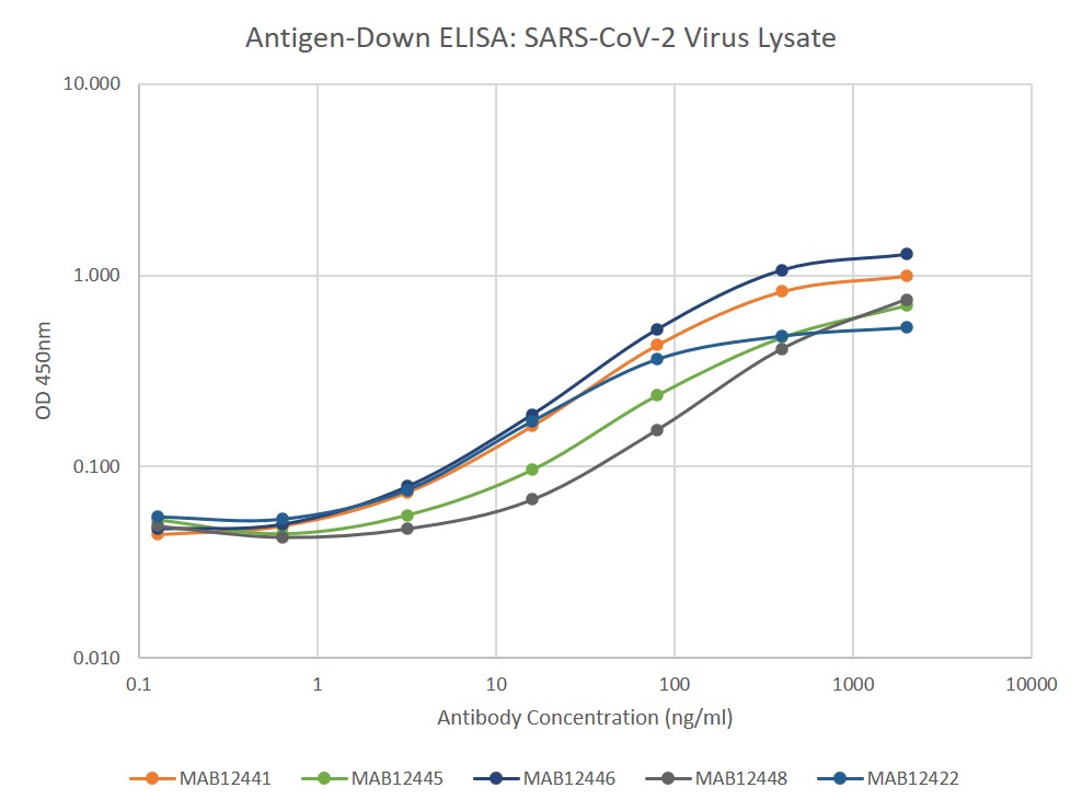
ELISA: Antigen-down ELISA showing binding of MAB12445 to immobilised SARS-CoV-2 virus lysate (NAT41605).
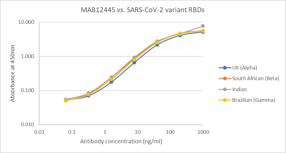
Direct ELISA: Plate was coated with 100µl of variant RBD proteins (UK, REC31946; South African, REC31945; Indian, REC31971; Brazilian, REC31961), at 1µg/ml and then incubated with 100µl MAB12445 antibody, diluted from 1000ng/ml to 0.064ng/ml. Diluted secondary IgG HRP antibody (100µl at 1:10,000) was then added. 100µl of TMB substrate (M0701A) was added in all wells and the reaction stopped after 10 min. with 1M HCl and the plate read at 405/450nm.
Mouse Anti SARS-CoV-2 Spike (S1) RBD Antibody (FG11)
Price range: £476.02 through £1,429.39 excl. VAT
Mouse anti SARS-CoV-2 spike (S1) RBD antibody (FG11) is a monoclonal antibody that is specific for the spike receptor binding domain of SARS-CoV and SARS-CoV-2, recognizing RBD from Wuhan-Hu-1, UK (Alpha), South African (Beta) Brazilian (Gamma) and Indian variants. No cross-reactivity observed in ELISA with SARS-CoV-2 subunit 2 (S2) or spike subunit 1 (S1) proteins from MERS-CoV, HCoV-NL63, HCoV-OC43, HCoV-229E and HCoV-HKU1. This antibody has been manufactured for use in ELISA immunoassay development.
MOUSE ANTI-SARS-COV-2 SPIKE (S1) RBD ANTIBODY (FG11)
Mouse anti SARS-CoV-2 spike (S1) RBD antibody (FG11) is a monoclonal antibody that is specific for the spike receptor binding domain of SARS-CoV and SARS-CoV-2, recognizing RBD from Wuhan-Hu-1, UK (Alpha), South African (Beta) Brazilian (Gamma) and Indian variants. No cross-reactivity observed in ELISA with SARS-CoV-2 subunit 2 (S2) or spike subunit 1 (S1) proteins from MERS-CoV, HCoV-NL63, HCoV-OC43, HCoV-229E and HCoV-HKU1. This antibody has been manufactured for use in ELISA immunoassay development.
PRODUCT DETAILS – MOUSE ANTI-SARS-COV-2 SPIKE (S1) RBD ANTIBODY (FG11)
- Antibody recognizes SARS-CoV and SARS-CoV-2 spike glycoprotein, subunit S1 receptor binding domain (RBD) for Wuhan-Hu-1, UK (Alpha), South African (Beta) Brazilian (Gamma) and Indian variants.
- No cross-reactivity in ELISA with SARS-CoV-2 spike subunit 2 (S2) or spike proteins from MERS-CoV, HCoV-NL63, HCoV-OC43, HCoV-229E and HCoV-HKU1.
- Isotype – Mouse IgG1
- Immunogen was recombinant SARS-CoV-2 spike RBD protein (REC31882, aa 1-223) expressed in HEK293 cells.
- Protein G purified from hybridoma culture supernatant.
- Suitable for use in ELISA, WB.
BACKGROUND
The severe acute respiratory syndrome coronavirus 2 (SARS-CoV-2) is the causative agent of the coronavirus induced disease 19 (COVID-19) which emerged in China in late 2019, resulting in a worldwide epidemic (Zhou et al., 2020). SARS-CoV-2 is an enveloped positive-sense single-stranded RNA virus with a number of important structural proteins, including the envelope (E) protein, the membrane (M) protein, the spike (S) protein, and the nucleoprotein (N). The S protein assists in the attachment of the virus to the human cell and comprises intracellular, transmembrane, and extracellular regions. The extracellular region contains the S1 receptor binding subunit (RBD) and the S2 membrane fusion subunit. Generally, following SARS-CoV-2 infection, antibodies appear after 7–14 days and persist for weeks after viral clearance. The most commonly detected antibodies are against the N protein and the S protein. Coronavirus neutralizing antibodies primarily target the trimeric spike (S) glycoproteins on the viral surface (Wang et al., 2020) and can change the course of infection in an infected individual by supporting virus clearance or protecting an uninfected host that is exposed to the virus (Prabakaran et al., 2009). However, the antibody responses against SARS-CoV-2 remain poorly understood (Tang et al., 2020) and better understanding of how the viral coating triggers a healthy immune system’s recognition and neutralisation of the virus is critical for optimisation of diagnostic tests (Petherick, 2020). It has been suggested that spike RBD may be a better target than N for diagnostic tests (Rosadas et al., 2020). The Native Antigens monoclonal antibodies, which are specific for SARS-CoV-2, have been manufactured to meet the need for improved COVID-19 diagnostic assays.
REFERENCES
- Petherick A. Developing antibody tests for SARS-CoV-2. Lancet. 2020 Apr 4;395(10230):1101-1102.
- Prabakaran P, Zhu Z, Xiao X, Biragyn A, Dimitrov AS, Broder CC, Dimitrov DS. Potent human monoclonal antibodies against SARS CoV, Nipah and Hendra viruses. Expert Opin Biol Ther. 2009 Mar;9(3):355-68.
- Rosadas C, Randell P, Khan M, McClure MO, Tedder RS. Testing for responses to the wrong SARS-CoV-2 antigen? Lancet. 2020 Sep 5;396(10252):e23.
- Tang YW, Schmitz JE, Persing DH, Stratton CW. Laboratory Diagnosis of COVID-19: Current Issues and Challenges. J Clin Microbiol. 2020 May 26;58(6):e00512-20.
- Wang C, Li W, Drabek D, Okba NMA, van Haperen R, Osterhaus ADME, van Kuppeveld FJM, Haagmans BL, Grosveld F, Bosch BJ. A human monoclonal antibody blocking SARS-CoV-2 infection. Nat Commun. 2020 May 4;11(1):2251.
- Zalzala HH. Diagnosis of COVID-19: Facts and challenges. New Microbes New Infect. 2020 Sep 16:100761.
- Zhou P, Yang XL, Wang XG, Hu B, Zhang L, Zhang W, Si HR, Zhu Y, Li B, Huang CL, Chen HD, Chen J, Luo Y, Guo H, Jiang RD, Liu MQ, Chen Y, Shen XR, Wang X, Zheng XS, Zhao K, Chen QJ, Deng F, Liu LL, Yan B, Zhan FX, Wang YY, Xiao GF, Shi ZL. A pneumonia outbreak associated with a new coronavirus of probable bat origin. Nature. 2020 Mar;579(7798):270-273.