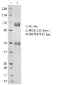
SDS-PAGE: Representative Coomassie-stained reducing SDS-PAGE showing purified Human T-Cell Lymphotropic Virus Type 1 (HTLV-1) envelope protein. Furin cleavage of the envelope protein occurs during the production process resulting in two bands. Both bands comprise the envelope protein as detected by western blot with anti-Fc tag.
Human T-Cell Lymphotropic Virus Type 1 (HTLV-1) Envelope Protein, Sheep Fc-Tag (HEK293)
$525.51 – $1,850.20 excl. VAT
Recombinant human T-cell lymphotropic virus type 1 (HTLV-1) envelope protein. The protein was produced in HEK293 cells and purified to high purity from culture supernatant with a TEV cleavage site and C-terminal sheep Fc-tag.
HUMAN T-CELL LYMPHOPTROPIC VIRUS TYPE 1 (HTLV-1) ENVELOPE PROTEIN, SHEEP FC-TAG (HEK293)
Recombinant human T-cell lymphotropic virus type 1 (HTLV-1) envelope protein. The protein was produced in HEK293 cells and purified from culture supernatant with a TEV cleavage site and C-terminal sheep Fc-tag.
PRODUCT DETAILS – HUMAN T-CELL LYMPHOPTROPIC VIRUS TYPE 1 (HTLV-1) ENVELOPE PROTEIN, SHEEP FC-TAG (HEK293)
- Human T-cell lymphotropic virus type 1 (HTLV-1) envelope protein (AA1-442).
- Strain Japan MT-2 subtype A.
- Produced in HEK293 cells and purified from culture supernatant.
- Purified by affinity chromatography, followed by dialysis.
- Presented in PBS at
BACKGROUND
HTLV-1 and HTLV-2 require cell-to-cell contact for efficient transmission and utilize envelope glycoprotein-mediated cell binding and entry (envelope glycoprotein gp62). Specific enzymatic cleavages in vivo yield mature proteins. Envelope glycoproteins are synthesized as an inactive precursor that is N-glycosylated and processed likely by host cell furin or by a furin-like protease in the Golgi to yield the mature SU (gp46) and TM (gp21) proteins. The cleavage site between SU and TM requires the minimal sequence [KR]-X-[KR]-R. HTLV-1 attachment and fusion to target cells begins when surface subunit (SU) of the HTLV-1 envelope glycoprotein interacts with three cellular surface receptors: Glucose Transporter (GLUT1), Heparin Sulfate Proteoglycan (HSPG) and the VEGF-165 receptor Neuropilin-1 (NRP-1). These receptors are widely distributed on target cells. The HTLV-1 and HTLV-2 surface (SU) and transmembrane (TM) subunits of Env share 65% and 79% residue identity, respectively. Despite this high similarity, HTLV-1 and HTLV-2 utilize a slightly different complex of receptor molecules. HTLV-1 utilizes heparan sulfate proteoglycan (HSPG) and neuropilin-1 (NRP1) for binding and glucose transporter 1 (GLUT1) for entry. HTLV-2 also utilizes NRP1 and GLUT1, but not HSPGs (Eusebio-Ponce et al., 2019; Martinez et al., 2019). The surface protein (SU) attaches the virus to the host cell by binding to its receptor. This interaction triggers the refolding of the transmembrane protein (TM) and is thought to activate its fusogenic potential by unmasking its fusion peptide. Fusion occurs at the host cell plasma membrane and the transmembrane protein (TM) acts as a class I viral fusion protein. Membrane fusion leads to delivery of the nucleocapsid into the cytoplasm.
REFERENCES
- Eusebio-Ponce E, Anguita E, Paulino-Ramirez R, Candel FJ. HTLV-1 infection: An emerging risk. Pathogenesis, epidemiology, diagnosis and associated diseases. Rev Esp Quimioter. 2019 Dec;32(6):485-496.
- Martinez MP, Al-Saleem J, Green PL. Comparative virology of HTLV-1 and HTLV-2. Retrovirology. 2019 Aug 7;16(1):21.


