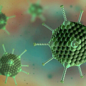
Canine Adenovirus
Canine adenovirus type 1 (Canine mastadenovirus A) is the causative agent of infectious canine hepatitis (ICH), a worldwide, contagious disease of dogs. ICH varies from a slight fever and congestion of the mucous membranes to severe depression, marked leukopenia, and coagulation disorders. It also is seen in foxes, wolves, coyotes, bears, lynx, and some pinnipeds (seals, sea lions, and walruses); other carnivores may become infected without developing clinical illness. Canine adenovirus type 2 is related to canine adenovirus type 1 and used in vaccines to provide protection against canine infectious hepatitis. It is also one of the causes of infectious tracheobronchitis, also known as canine cough (kennel cough). ICH has become uncommon in areas where routine immunization is carried out, but periodic outbreaks, which may reflect maintenance of the disease in wild and feral hosts, reinforce the need for continued vaccination.
Canine Adenovirus Background
Both canine adenovirus (CAdV-1) and canine adenovirus 2 (CAdV-2) are spread similarly, but the resulting diseases are vastly different. CAdV-1 is the causative agent of infectious canine hepatitis (so-called Rubarth’s disease), a life-threatening disease of puppies. It also is seen in foxes, wolves, coyotes, jackals, otters, raccoons, skunks, bears, lynx, and some pinnipeds (Seals, Sea Lions, and Walruses). CAdV-2 is serologically indistinguishable from CAdV-1 and causes a localized respiratory disease in dogs; it is one of the causes of infectious canine tracheobronchitis (kennel cough) and is often found in dog breeder stocks.
Both adenoviruses are spread from dog-to-dog interactions through infected respiratory secretions or contact with contaminated feces or urine. Ingestion of urine, feces, or saliva of infected dogs is the main route. Recovered dogs shed virus in their urine for at least 6 months. Initial infection occurs in the tonsillar crypts and Peyer patches, followed by viremia and disseminated infection. Vascular endothelial cells are the primary target, with hepatic and renal parenchyma, spleen, and lungs becoming infected as well. Chronic kidney lesions and corneal clouding (blue eye) result from immune-complex reactions after recovery from acute or subclinical disease. Respiratory disease in affected dogs is characterized principally by bronchitis and bronchiolitis. An essential difference between canine adenoviruses 1 and 2 is that, whereas canine adenovirus 1 causes systemic disease, canine adenovirus 2 infection results only in restricted respiratory disease. The molecular basis of this difference remains uncertain, but this property is exploited for vaccination of dogs: specifically, although the use of live-attenuated canine adenovirus 1 vaccines sometimes results in blue eye because of the ability of the vaccine virus to replicate systemically, canine adenovirus 2 vaccines do not replicate systemically. Canine adenovirus 2 vaccines, however, provide complete homologous and cross-protection against disease induced by canine adenovirus 1. Whereas canine adenovirus 1 is a common infection of foxes, wolves, and coyotes, evidence for canine adenovirus 2 infections in wildlife is lacking.
Most infections with canine adenovirus 1 are asymptomatic, a situation probably enhanced by the introduction of vaccination. Attenuated live canine adenovirus 2 vaccines are available and the antigenic relationship between canine adenoviruses 1 and 2 is sufficiently close for the canine adenovirus 2 vaccine to be cross-protective; it also has the advantage that it does not cause corneal edema or ‘blue eyes’.
References
- Fenner’s Veterinary Virology (Fifth Edition) 2017, Chapter 10 – Adenoviridae. Pages 217-227.
Canine Adenovirus Antigens
The Native Antigen Company is pleased to offer antigens for both Canine Adenovirus 1 and Canine Adenovirus 2, which can be used for immunoassay development and other applications.
Questions?
Check out our FAQ section for answers to the most frequently asked questions about our website and company.


