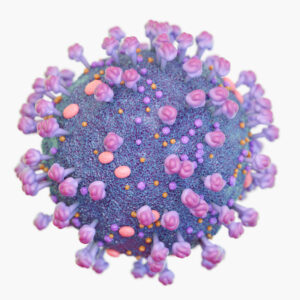
Feline Immunodeficiency Virus
Feline Immunodeficiency Virus Background
FIV compromises the immune system of cats by infecting many cell types, including CD4+ and CD8+ T lymphocytes, B lymphocytes, and macrophages. The virus gains entry to the host’s cells through the interaction of the envelope glycoproteins of the virus and the target cells’ surface receptors. In contrast to human immunodeficiency virus (HIV), FIV uses CD134 instead of CD4 as its primary receptor. CD134, which is also known as OX40, is a member of the tumour necrosis factor and nerve growth factor receptor superfamily (Taniwaki et al., 2013). Binding of the viral envelope glycoprotein (SU) and CD134 changes the shape of the SU protein to facilitate interaction between SU and the chemokine receptor CXCR4. This interaction causes the viral and cellular membranes to fuse, allowing the transfer of the viral RNA into the cytoplasm, where it is reverse transcribed (into DNA provirus) and integrated into the cellular genome through non homologous recombination, resulting in lifelong infection. The virus can lay dormant in the asymptomatic stage for long periods of time without being detected by the immune system or can cause lysis of the cell.
FIV contains a diploid genome which consists of two identical single-strands of RNA (~9400 nt) in positive-sense orientation flanked by Long Terminal Repeats (LTR) and contains the characteristic retroviral genes gag, pol, and env. The FIV genome also encodes the regulatory proteins Rev, Vif, and OrfA, but lacks the accessory HIV-1 genes vpr, vpu, and nef, as well as tat, whose product regulates viral genome transcription. The FIV Gag polyprotein contains all the necessary information for virion assembly and budding. It is cleaved into matrix (MA), capsid (CA) and nucleocapsid (NC) proteins. The capsid protein derived from the polyprotein Gag is assembled into a viral core (the protein shell of a virus) and the matrix protein also derived from Gag forms a shell immediately inside of the lipid bilayer. The Env polyprotein encodes the surface glycoprotein (SU) and transmembrane glycoprotein (TM). Both SU and TM glycoproteins are heavily glycosylated, which may mask the B-cell epitopes of the Env glycoprotein giving the virus resistance to neutralizing antibodies. FIV assembly occurs at the plasma membrane of the infected cells as the result of the multimerization of the Gag polyprotein into virions, which are then released into the extracellular medium (González & Affranchino, 2018).
FIV infection is one of the most important infectious diseases in cats because it causes persistent infection (Taniwaki et al., 2013). It also serves as a useful model for HIV studies (Bendinelli et al., 1995). However, as with HIV, the development of an effective vaccine against FIV is difficult because of the high number and variations of the virus strains.
References
- Westman et al. (2019). Diagnosing feline immunodeficiency virus (FIV) and feline leukaemia virus (FeLV) infection: an update for clinicians. Australian Veterinary Journal Volume 97 No 3, 47-55.
- Bendinelli et al. (1995). Feline Immunodeficiency Virus: an Interesting Model for AIDS Studies and an Important Cat Pathogen. Clinical Microbiology Reviews. 87–112.
- González & Affranchino (2018). Properties and Functions of Feline Immunodeficiency Virus Gag Domains in Virion Assembly and Budding. Viruses. 10(5): 261.
- Taniwaki et al. (2013). Virus–host interaction in feline immunodeficiency virus (FIV) infection. Comp Immunol Microbiol Infect Dis. 36(6):549-57.
Feline Immunodeficiency Virus Antigens
Questions?
Check out our FAQ section for answers to the most frequently asked questions about our website and company.

