Human Parainfluenza Virus
Human Parainfluenza Virus Background
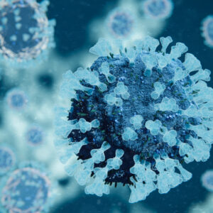
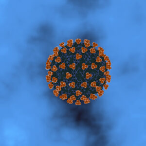
HPIV virions contain a single molecule of linear, negative sense, ssRNA that is not infectious alone but is infectious in the form of the nucleocapsid. The RNA genome size varies and is 15,600 nt for human parainfluenza virus 1 (HPIV1), 15,654 nt for human parainfluenza virus 2 (HPIV-2), 15,462 nt for human parainfluenza virus 3 (HPIV3) and 15,246 nt for parainfluenza virus 5 (PIV5, previously known as simian virus 5 [SV-5]) (ICTV, 2011). HPIV are enveloped and of medium size (150 to 250 nm). The single, negative-sense RNA strand encodes six essential proteins in a conserved order: the nucleocapsid protein (NP), the phosphoprotein (P), the matrix protein (M), the fusion glycoprotein (F), the hemagglutinin neuraminidase (HN) glycoprotein, and the RNA polymerase (L). The HN and fusion glycoproteins are surface proteins which mediate attachment to the sialic acid residues on the surface of host epithelial cells (HN) and fusion of the viral envelope with the host cell membrane (F), respectively. The HN protein also facilitates release of new virions from the cell by cleaving the sialic acid residue (reviewed in Branche & Falsey, 2016).
Parainfluenza virus infections occur throughout the world with seasonal variations in serotype-specific rates of infection which is determined by region. HPIV3 is the most commonly isolated serotype in symptomatic disease for both children and adults. However, during epidemic years, HPIV1 is associated with significant disease burden and hospitalizations in children (reviewed in Branche & Falsey, 2016). Among non-immune compromised adults, HPIV infection typically causes mild disease manifested as upper respiratory tract symptoms and is infrequently associated with severe croup or pneumonia. HPIV infection may also be associated with viral exacerbations of chronic airway diseases, asthma or COPD or chronic rhinosinusitis. After respiratory syncytial virus, HPIVs are the second most common cause of acute respiratory tract infections among children aged less than 5 years, possibly accounting for up to 17% of hospitalizations. Serological surveys have indicated that 60% of the children are infected with HPIV-3 by 2 years of age, and this number increases up to 80% by 4 years of age (reviewed in Pawełczyk & Kowalski, 2017).
HPIVs usually spread from an infected person to others through the air by coughing and sneezing, close personal contact and contaminated surfaces. Infection begins in the nose and oropharynx and then spreads to the lower airways with peak replication 2 to 5 days after initial infection. Host defense against HPIV is mediated by both humoral and cellular immunity. Serum antibodies directed against the two surface glycoproteins, F and HN, are neutralizing and protective against challenge. Neutralizing antibody appears to be serotype specific with little cross protection afforded by antibodies between HPIV serotypes 1 to 4. Cytotoxic T lymphocyte responses are important for clearance of virus, and T cell epitopes have been demonstrated on the HN, P, and NP proteins of HPIV. Repeated infections are often needed to fully protect a child’s lower respiratory tract from HPIV infection and eventual protection may be a combination of high levels of neutralizing antibody and cellular immunity. Immunity to HPIV is incomplete and reinfections with any of the HPIV serotypes can occur throughout life (reviewed in Branche & Falsey, 2016).
Currently, there is no licensed vaccine for the prevention of parainfluenza infection and no specific antiviral treatment for HPIV illness. The HPIV hemagglutinin and neuramindase proteins are more stable compared with those of influenza A viruses, but antigenic differences have been noted over time, producing strains serologically and genetically different from earlier isolates and impeding vaccine development (reviewed in Branche & Falsey, 2016).
References
- Branche AR, Falsey AR. Parainfluenza Virus Infection. Semin Respir Crit Care Med. 2016;37(4):538-554.
- Centers for Disease Control and Prevention (2019). Human Parainfluenza Viruses (HPIVs).
- ICTV 9th Report (2011). Paramyxoviridae.
- Henrickson, KJ. Parainfluenza Viruses. Clin Microbiol Rev. 2003 Apr; 16(2): 242–264.
- Pawełczyk M, Kowalski ML. The Role of Human Parainfluenza Virus Infections in the Immunopathology of the Respiratory Tract. Curr Allergy Asthma Rep. 2017;17(3):16.
- Xu R, Palmer SG, Porotto M, et al. Interaction between the hemagglutinin-neuraminidase and fusion glycoproteins of human parainfluenza virus type III regulates viral growth in vivo. mBio. 2013;4(5):e00803-e813.
Human Parainfluenza Virus Antigens

Human Parainfluenza Virus Type 3 Lysate
Price range: $845.03 through $3,594.50 excl. VAT![Influenza B [B/Palermo/4/2013] Hemagglutinin Parainfluenza Virus Type 4A](https://thenativeantigencompany.com/wp-content/uploads/2020/12/Coronavirus-35-300x300.jpg)
Human Parainfluenza Virus Type 4A Lysate
Price range: $845.03 through $3,594.50 excl. VAT
Human parainfluenzavirus 3 haemagglutinin-neuraminidase (HN) protein, C-terminal His-tag
Price range: $915.56 through $3,890.19 excl. VAT
Questions?
Check out our FAQ section for answers to the most frequently asked questions about our website and company.
