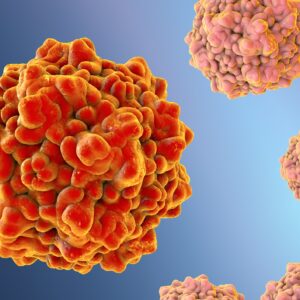
Porcine Parvovirus
Porcine parvovirus (PPV, Protoparvovirus genus) is a small non-enveloped virus and is considered to be one of the major causes of porcine reproductive failure. PPV normally multiplies in the intestine of the pig without causing clinical signs; however, the virus can cross the placental barrier during the infection and is characterized by embryonic and fetal infection and death. PPV is ubiquitous in pig populations world-wide and is one of the most common and important infectious agents of infertility.
Porcine Parvovirus Background
Currently PPV is classified as the Protoparvovirus genus (species Ungulate protoparvovirus 1). In recent years many novel porcine parvoviruses have been described such as porcine parvovirus 2 (PPV2) and porcine parvovirus 3 (PPV3) and these have been classified as Tetraparvovirus genus (species Ungulate tetraparvovirus 3 and Ungulate tetraparvovirus 2, respectively), porcine parvovirus 4 (PPV4) into the Copiparvovirus genus (species Ungulate copiparvovirus 2). Porcine parvovirus 6 (PPV6) is described by ICTV as a related, unclassified virus in the Copiparvovirus genus. Porcine parvovirus 5 (PPV5) is still not classified in the ICTV taxonomy proposal, but genetic analysis showed that this virus is most closely related to PPV4. The most recently discovered porcine parvovirus 7 (PPV7) is also unclassified, but in contrast to PPV5, it is only distantly related to the other porcine parvoviruses, and for this virus, the new genus Chappaparvovirus was proposed (Miłek et al., 2019).
Porcine parvovirus was first found in primary cell cultures from porcine kidney and testicle used to cultivate hog cholera virus in 1965 in Munich, Germany, by Anton Mayr and coworkers. They were observed as persistent contaminant small particles (22–23 nm size). The particles were similar to the Kilham rat virus (a parvovirus) and due to the replication ability of the virus in cell lines from swine, it was isolated and classified as a porcine parvovirus. The genome of parvoviruses comprise a single stranded DNA molecule of about 5 kb. The genome of PPV encodes four proteins and uses alternative splicing to extend the coding capacity. Two nonstructural proteins, NS1 and NS2, operate in the replication of the virus, particularly for DNA replication. Two structural proteins (VP1 and VP2) are transcribed and translated from the parvovirus genome (Streck & Truyen, 2019).
The major clinical sign of PPV infections in pigs is maternal reproductive failure and was first described by Cartwright and Huck in 1967. Porcine parvovirus infection (PPV) is now recognized as a common and important cause of infectious infertility. PPV is one of the organisms listed as responsible within the Stillbirths Mummification Embryonic Death and Infertility (SMEDI) syndrome. Porcine parvoviral infection (PPV) is usually subclinical and a very common infection. Prior to porcine reproductive and respiratory syndrome (PRRS), PPV was probably the most commonly diagnosed infectious cause of reproductive failure in swine (Mészáros et al., 2017).
PPV is most often transmitted either by mouth or through the snout, passing into the intestine where it multiplies and subsequently passed out in faeces. The primary replication of the PPV occur in lymphoid tissues. Following infection, virus is shed in the secretions and excretions from the animal for approximately two weeks. Porcine parvovirus shows high stability in the environment and can remain infectious for months; contaminated instruments or stables may therefore be a constant source of infection. The virus can be transported between herds via fomites (e.g. clothes, boots and equipment) and it is also speculated that infected boars can introduce the virus into new herds. It is also resistant to most disinfectants, and these greatly contribute to why it is widespread and difficult to eliminate.
Currently, PPV vaccines represent cell culture derived virus (usually the non-pathogenic NADL-2 strain) which is chemically inactivated (by formalin, beta-propiolactone or binary ethyleneimine), mixed with oil or aluminium hydroxide as adjuvants and administered parenterally. The use of these vaccines induces antibody titres that can reduce clinical manifestations, but cannot prevent infection. There is no specific treatment for parvovirosis and general management measures aimed at promoting good health status of the herd are the best control. It has been observed that new mutations in the capsid protein can modify antigenic properties, and may reduce the cross neutralization ability of sera raised against strains commonly used in commercial vaccines. Therefore, new vaccine formulations that induce long-lasting immunity and cover all the dominant virus strains circulating in pig populations are likely to be needed in the future (Streck & Truyen, 2019).
References
- Streck AF, Truyen U. Porcine Parvovirus. Curr Issues Mol Biol. 2020;37:33-46.
- Mészáros I, Olasz F, Cságola A, Tijssen P, Zádori Z. Biology of Porcine Parvovirus (Ungulate parvovirus 1). Viruses. 2017 Dec 20;9(12):393.
- Miłek D, Woźniak A, Guzowska M, Stadejek T. Detection Patterns of Porcine Parvovirus (PPV) and Novel Porcine Parvoviruses 2 through 6 (PPV2-PPV6) in Polish Swine Farms. Viruses. 2019 May 24;11(5):474.
Porcine Parvovirus Antigens
The Native Antigen Company is pleased to offer a recombinant porcine parvovirus 2 (PPV2) capsid 1 protein, manufactured in insect cells to high purity for assay development and research applications.
Questions?
Check out our FAQ section for answers to the most frequently asked questions about our website and company.

