Rift Valley Fever
Rift Valley fever (RVF) is an acute, fever-inducing viral disease, most commonly observed in domesticated animals (such as cattle, buffalo, sheep, goats, and camels), with the ability to infect and cause illness in humans. RVF is generally found in regions of eastern and southern Africa where sheep and cattle are raised, though the virus exists in most of sub-Saharan Africa, including west Africa and Madagascar. In September 2000, an RVF outbreak was reported in Saudi Arabia and subsequently, Yemen. This outbreak represents the first instance of Rift Valley fever identified outside of Africa.
The Native Antigen Company offers mammalian-expressed RVFV antigens for use in research and assay development for diagnostic testing.
Rift Valley Fever Virus Background
Rift Valley fever is an emerging zoonotic, mosquito-borne viral disease that primarily affects livestock but can also affect humans. RVF causes significant loss to livestock and severe illness and death in humans, resulting in significant economic impact to countries where RVF is prevalent.
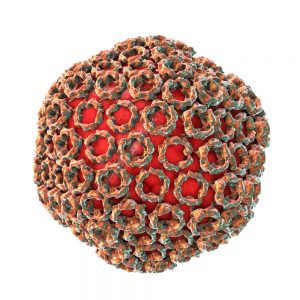
RVF is caused by the Rift Valley fever virus (RVFV), which was first recognised in the 1930s, in the Rift Valley region of Kenya. Rift Valley fever virus is an enveloped RNA virus that belongs to the genus Phlebovirus, a member of the Bunyaviridae family. RVFV is an arthropod-borne virus that is transmitted to domesticated livestock by mosquitoes. Several species of mosquito transmit RVFV and the vector varies according to geographical region. Cattle, sheep, goats and camels are particularly susceptible to RVF and serve as amplifying hosts for the virus. Reports of RVFV outbreaks outside sub-Saharan Africa have raised concerns that RVFV may spread to temperate climates through emerging, competent vectors.
Humans may become infected through contact or via open wounds, blood products, organs and bodily fluids from animals infected with RVFV. In some cases, humans may be infected through the bite of infected mosquitoes, but this is uncommon. Human to human transmission of RVFV has not been reported to date (Pepin M, et al).
In humans, the incubation period of RVFV is 2-6 days and infection may present in different ways. In many cases, individuals remain asymptomatic or may present with mild fever, arthralgia, myalgia and headaches. In some cases, patients have clinical symptoms that can be confused with meningitis. A small percentage of patients develop severe forms of RVF disease, which may present as ocular disease, meningoencephalitis or haemorrhagic fever. The haemorrhagic form of RVF carries the highest mortality risk (CDC).
Currently, there is no antiviral therapy for the treatment of symptomatic RVF, and there is no licensed prophylactic vaccine to prevent RVFV infection. Diagnosis of RVFV in humans is achieved using serological methods, reverse transcriptase polymerase chain reaction (RT-PCR) or cell culture to isolate the virus. In 2015, the World Health Organization identified RVFV as an emerging disease that is likely to cause a severe epidemic and a significant risk to public health (WHO). Recent outbreaks of RVFV in Gambia and Kenya have renewed concerns about this emerging virus.
References
- Pepin M et al (2010). Rift Valley fever virus (Bunyaviridae: Phlebovirus): an update on pathogenesis, molecular epidemiology, vectors, diagnostics and prevention. Vet Res.41(6):61.
- Centers for Disease Control and Prevention: Rift Valley fever (RFV)
- The World Health Organization: Factsheet, Rift Valley fever
Rift Valley Fever Antigens
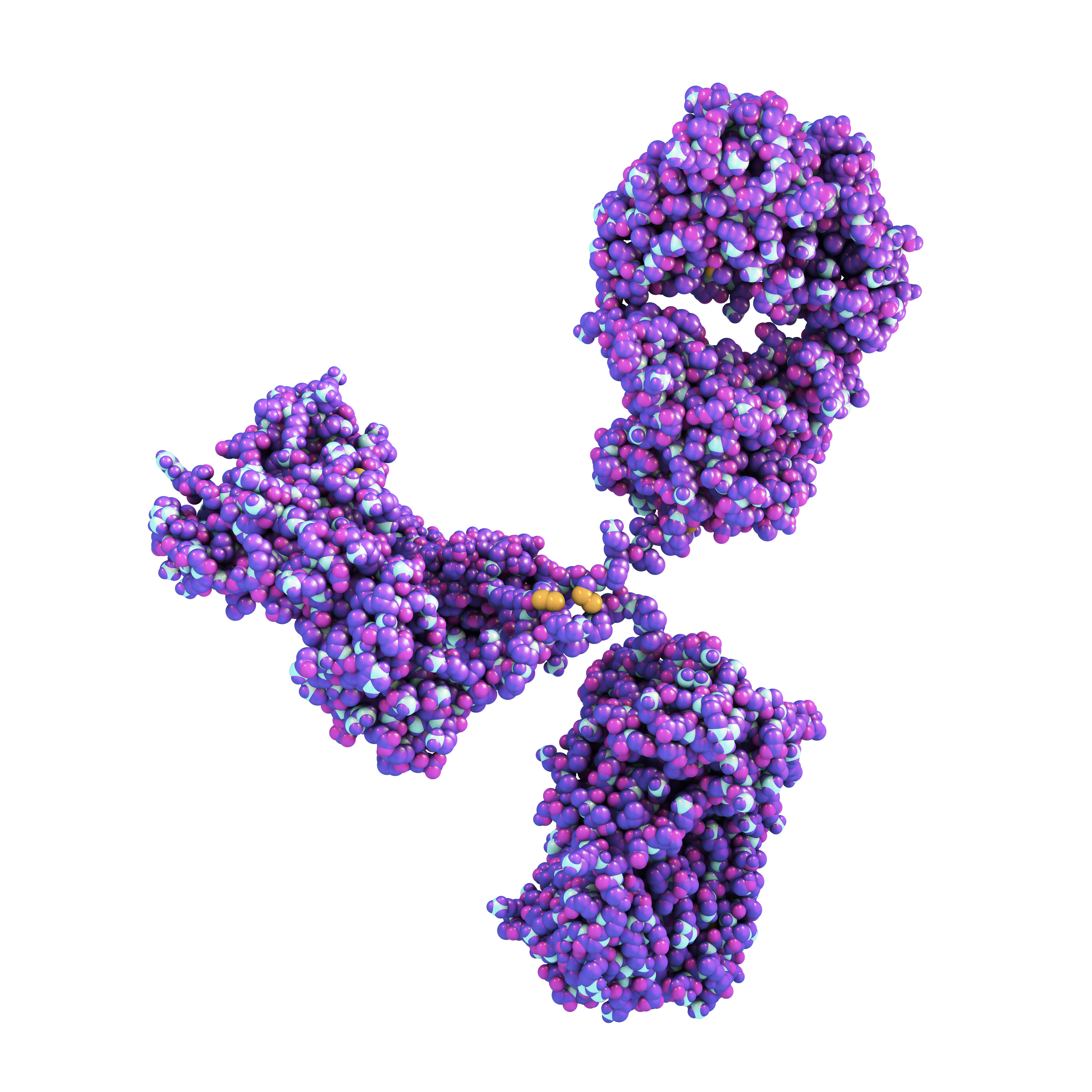
Rift Valley Fever Virus Nucleoprotein has been manufactured in response to the unmet need for highly purified, concentrated protein, for use in serological-based diagnostic assays. RVFV nucleoprotein is engineered in a human cell line, using state-of-the-art expression and purification techniques. The protein comprises residues 2-245, linked to an N-terminal His-tag, via a 10 amino acid glycine-serine linker.

Mouse Anti-Rift Valley Fever Virus Nucleoprotein (M975)
Price range: $454.04 through $1,139.31 excl. VAT
Mouse Anti-Rift Valley Fever Virus Nucleoprotein (M976)
Price range: $454.04 through $1,139.31 excl. VAT
Mouse Anti-Rift Valley Fever Virus Nucleoprotein (M977)
Price range: $454.04 through $1,139.31 excl. VAT
Rift Valley Fever Antibodies
Rift Valley Fever Virus antibodies are specific for RVFV nucleoprotein and suitable for use in ELISA and western blot.
Questions?
Check out our FAQ section for answers to the most frequently asked questions about our website and company.

